
Acquired stenosis of the external auditory canal (EAC) is uncommon,1 with several related causes. However, no reports exist of acquired stenosis of EAC secondary to paraneoplastic syndrome. This is the first report of auricular pyoderma gangrenosum manifesting as paraneoplastic syndrome of renal clear cell carcinoma, evolving to stenosis of the EAC.
Case reportMale, 56, presenting with an ulceration on the right auricular concha for 45 days, with poor response to topical and systemic antibiotics, associated with otorrhea, hearing loss, otalgia and fever. Otoscopy revealed an ulcerated lesion in the auricle and edema of the EAC, preventing appropriate visibilization of the tympanic membrane.
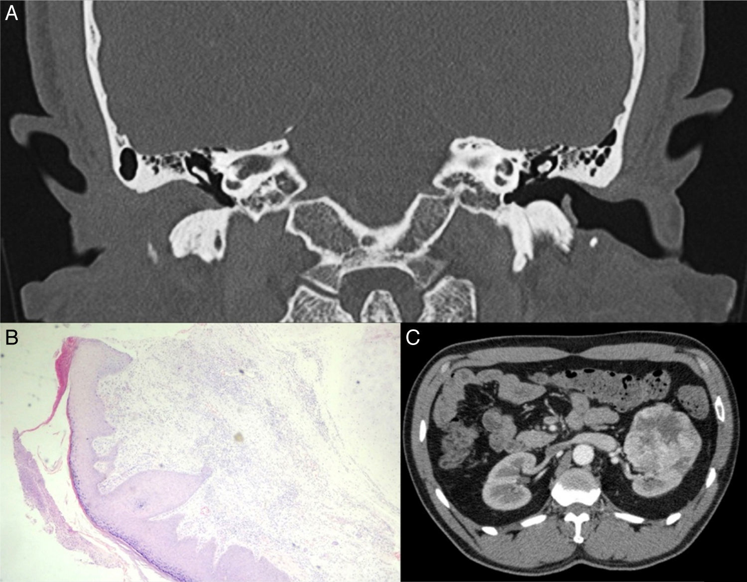
Computed tomography (CT) of the temporal bones (Fig. 1A) showed obliteration of the EAC without regional bone involvement. Due to the possibility of malignant external otitis, the patient was treated with Piperacillin and Tazobactam for 21 days. The lesion improved after two weeks, evolving to scar stenosis of the EAC. The patient was discharged after 21 days with antibiotic therapy (Ciprofloxacin 750mg, 21 days) and office follow-up for possible surgery for stenosis correction.
 Temporal bone tomography (CT) showing obliteration of right EAC. (B) Photomicrograph of the external ear biopsy suggestive of pyoderma gangrenosum (Hematoxylin and eosin staining; 10×). (C) Contrast abdominal CT showing the tumor in the left kidney.")
After 3 months, the patient presented with severe bilateral otalgia associated with otorrhea, and necrosis of the auricular concha, tragus and intertragic incisure bilaterally. Histopathologic examination revealed nonspecific chronic inflammation affecting the dermis and the perichondrium with focal ulceration of the skin, suggestive of pyoderma gangrenosum (Fig. 1B). Multisensible Acinetobacter baumani was isolated in the culture. Inflammatory markers, C3, C4, p-ANCA, c-ANCA and ANA were normal.
An isodense mass was observed in an abdominal CT, located in the middle third of the left kidney, heterogeneous, with peripheral contrast uptake of 6cm×6.4cm×6cm (Fig. 1C). The patient subsequently underwent a left nephrectomy with histological diagnosis of a clear cell variant of renal carcinoma.
DiscussionStenosis of the EAC is rare, with an incidence of 0.6 cases per 100,000 inhabitants.2 The most common cause is chronic external otitis. However, recurrent or persistent suppurative otitis media, foreign body, trauma, radiation, tumors are also reported.1
Chronic ulcerated lesions affecting the EAC and auricles can be secondary to several diseases, including malignant external otitis, skin infection (fungal, mycobacterial or viral), insect bites, lymphoma, cutaneous primary tumor, metastasis, perichondritis, vasculitis, Wegner's granulomatosis, artifact dermatitis, neurodermatitis and pyoderma gangrenosum.3
In 50% of cases, pyoderma gangrenosum (PG) is associated with neoplasm, illicit drugs, systemic inflammatory diseases (ulcerative colitis, Behcet's disease, Crohn's disease, Wegner's granulomatosis and rheumatoid arthritis) and myeloproliferative diseases.4 The pathogenesis of PG remains unclear, but a dysfunction of neutrophil chemotaxis might be involved.3,5
PG is a diagnosis of exclusion as the histopathologic findings are not specific. A biopsy including the border of the ulcer and the adjacent skin is necessary to exclude vasculitis, malignancy and infection. Histologic features of the untreated ulcer usually include dense neutrophilic dermal infiltrates and necrosis of the overlying epidermis.6 Investigation of the underlying cause is performed with laboratory exams, such as serum protein electrophoresis, anti-nuclear antibodies and anti-neutrophil cytoplasmatic antibodies, and colonoscopy.3
The clear cell renal carcinoma is the third most common type of neoplasm in the genitourinary system.7 Paraneoplastic syndrome is the initial manifestation in about 40% of cases.8 This is the first described case of pyoderma gangrenosum as paraneoplastic syndrome related to this type of neoplasm.9
This patient presented with no other signs suggesting systemic involvement. In view of a negative serology and inflammatory markers and disease recurrence after empirical treatment of malignant external otitis, imaging tests were performed to exclude relapsing polychondritis and neoplasms. With total gross removal of the tumor, there was no recurrence.
Final commentsAcquired EAC stenosis is uncommon. It is important to diagnose the underlying related disease. Among the disease possibilities, the paraneoplastic disease should be considered.
Conflicts of interestThe authors declare no conflicts of interest.
Please cite this article as: Falqueto LE, Kaddoum ML, Miranda MM, Ramos HF. Acquired stenosis of external auditory canal secondary to paraneoplastic manifestation of renal cancer. Braz J Otorhinolaryngol. 2018;84:249–51.
Peer Review under the responsibility of Associação Brasileira de Otorrinolaringologia e Cirurgia Cérvico-Facial.
gology is pleased to honor the reviewers


