
Lacrimal sac mucocele (LSM) is characterized by obstruction of the nasolacrimal duct (NLD) with consequent dilatation and distension of the lacrimal sac (LS) by mucopurulent material.1 This report aimed to describe this condition, which is rare in adults, discussing its formation mechanisms, differential diagnosis, and treatment.
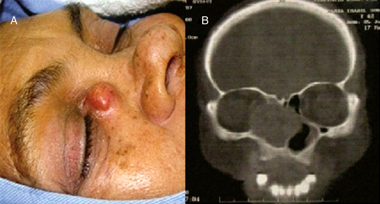
Case reportA female patient, 54 years old, complained of excessive tearing in the right eye for 12 years. After four years, she presented with progressive obstruction of the nasal passages. Approximately one year before presenting to this service, a bulging swelling, painful upon compression, appeared near the inner corner of the right eye. On physical examination, she demonstrated epiphora in the right eye and a cystic lesion associated with redness of the overlying skin, close to the medial corner of the right eye, measuring 1.2cm in diameter (Fig. 1A).
 Macroscopic aspect of the lesion in the medial region to the eyeball. (B) Computed tomography of the paranasal sinuses, coronal section.")
The swelling showed no drainage on expression, nor regurgitation of secretion through the canaliculi. Anterior rhinoscopy disclosed total obstruction of the right nasal cavity and, on the left, reduced lumen due to septal deviation. Computed tomography (CT) evidenced a lesion with soft tissue consistency at the ethmoid, right maxillary sinus, and nasal cavity, showing erosion of the lamina papyracea, with compression of the medial rectus muscle and eyeball displacement, as well as erosion and displacement of the nasal septum to the left, consistent with mucocele (Fig. 1B). Thus, surgical treatment was performed, which disclosed a pouch filled with mucopurulent material originating from the lacrimal sac. Surgical marsupialization was carried out. After six months, the patient remains asymptomatic.
DiscussionLSM rarely affects adults.2 It is caused by the accumulation of tear secretion in the medial canthal region due to obstruction of the nasolacrimal duct, generating a dacryocystocele, followed by the appearance of an infectious/inflammatory process that characterizes the mucocele.3,4 On physical examination, the characteristic of LSM is a non-compressible mass in the medial canthal region, with or without associated cellulitis of the overlying skin.1,3
In adults, LSM is often a result of acquired chronic obstruction of the NLD1,2 and secondary blockage of canaliculi. The NLD obstruction may be due to chronic infections, mainly low pathogenicity bacteria and fungi; dacryoliths anatomical alterations, such as anomalous ethmoid cells, facial fractures, or complications of nasal surgeries; and use of certain drugs (fluorouracil and docetaxel).1,5 Neoplasms of the nasolacrimal sac or duct or of adjacent structures are extremely rare causes of NLD obstruction. Acquired NLD obstruction is more common in females.6
The erosion of facial bones resulting from a long-term compression is mediated by the production of inflammatory mediators or compressive effect.2
It is essential to obtain a CT of the paranasal sinuses if other LS pathologies or significant nasal comorbidities are suspected, or for preoperative review.2 Magnetic resonance imaging is valuable in the diagnosis of LS tumors, which are usually malignant and can mimic mucoceles on CT scans.2
The differential diagnosis of LSM should include dacryocystitis, lacrimal sac diverticulum, encephalocele, ethmoid and maxillary mucoceles, dermoid or epidermoid cysts, and neoplasia of the LS or contiguous structures.1–6
Neoplasia is suspected when the lesion occurs above the medial canthal ligament, if there is severe pain, bloody discharge through the lacrimal canaliculi, or palpable mass in the topography of LS and bone destruction.1–6
The treatment is commonly performed together by the ophthalmologist and otolaryngologist. In adults, the treatment of choice is dacryocystorhinostomy due to the ineffectiveness of conservative treatment and rarity of spontaneous regression.5 Satisfactory results have also been reported with stent placement in the nasolacrimal duct.3
Final commentsLSM in adults is most often a complication of chronic dacryocystitis caused by chronic obstruction of the NLD and secondary blockage of canaliculi. The evaluation of choice is computed tomography of the paranasal sinuses and the indicated treatment is dacryocystorhinostomy.
Conflicts of interestThe authors declare no conflicts of interest.
Please cite this article as: do Nascimento SB, Rodrigues AB, Jurity TP, de Sá JC, Castelo Branco AN. Lacrimal sac mucocele. Braz J Otorhinolaryngol. 2014;80:540–1.


