
Dental implants can be accidentally introduced into the maxillary sinus both during and after surgical installation. Both situations are uncommon and usually induce sinusitis or others important complications.1 The aim of the present study was to report a rare clinical case of late displacement of a dental implant into the maxillary sinus, in which a Caldwell-Luc (CL) approach was performed, followed by the reconstruction of the anterior maxillary sinus wall using a titanium mesh.
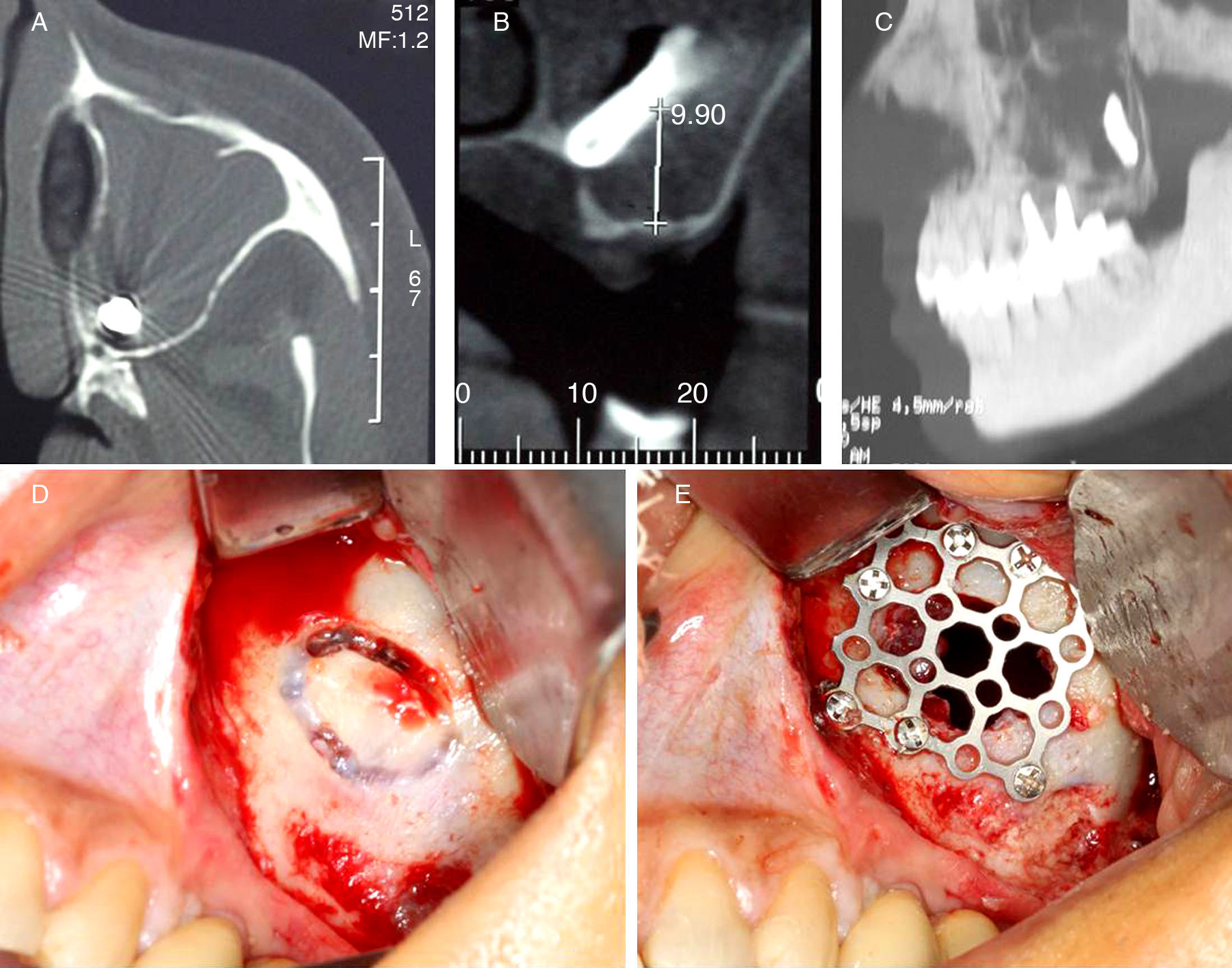
Case presentationA 49-year-old woman was treated six months previously with dental implants, and received three in the left posterior maxilla. She was referred to this clinic since one of the three implants disappeared during casting. CT scan showed what appeared to be a dental implant located posteriorly and inferiorly inside of the maxillary sinus (Fig. 1A–C). The mucosa from the sinus demonstrated important alterations; its density was compatible with acute sinusitis, corroborating the clinical findings, which included airway obstruction and moderate facial pain. There was no evidence of oro-antral fistula. After antibiotic therapy for acute sinusitis, the dental implant was removed through a CL approach. Under local anesthesia, a small surgical incision was made on the buccal sulcus. The anterior bony wall of the sinus was exposed, and an access was made with a carbide round bur (Fig. 1D). The sinus mucosa around the dental implant was resected, followed by intense cleaning. Finally, the anterior wall of maxillary sinus was reconstructed using a titanium mesh, which was fixated by monocortical screws (Fig. 1E). The patient has been followed up for 32 months, with no complications.
Discussion TC axial, (B) TC coronal, (C) TC sagittal, (D) Calwell-Luc approach and (E) reconstruction of the sinus anterior wall with a titanium mesh.")
Poor bone quality and quantity of the posterior maxilla, in addition to alveolar pneumatization from maxillary sinus, are predisposing factors for the displacement of dental implants. The major risk factor is inadequate surgical technique, which includes overtreatment of the implant preparation, sinus floor perforation, and poor primary stability. Late displacement is rare and usually happens during the first six months after implantation.2 In the present case, the displacement occurred after six months, during manipulation of the implant for prosthetic rehabilitation.
There are several methods to remove a dental implant from the maxillary sinus, such as suction through bone alveolar defect, CL approach, functional endoscopy sinus surgery (FESS), and transoral endoscopy approach via canine fossa.3,4 In the past few decades, FESS has been replacing the CL approach for the treatment of paranasal pathologies, since it has been more effective. Besides all its advantages, isolated FESS is not effective in removing larger materials, especially those located in the posterior and inferior aspects of the sinus.5,6 The presence of an oral–antral communication, inflammatory alterations of sinus mucosa, and ostium patency also must be taken into account to choose the correct plan of treatment. The anterior maxillary sinus wall reconstruction becomes important due to potential complications from the CL approach, such as persistent bone defect and retractions of the soft tissues of the cheek.5,6 Bone grafts, guided tissue regeneration, and buccal fat pad have been used to decrease CL failure.1 For this purpose, a titanium mesh was used in the present case.
Final remarksThe CL approach is still indicated to remove objects located posteriorly/inferiorly inside of the maxillary sinus, with additional care to reconstruct the bone defect created. Endoscopy procedures isolated or in association with the CL approach are also proven to be effective. The professional must take account these three alternatives in order to choose the most suitable procedure.
Conflicts of interestThe authors declare no conflicts of interest.
Please cite this article as: Tavares RN, Nogueira AS, Sampieri MB, Bezerra MF, Gonçales ES. Late displacement of a dental implant into maxillary sinus. Braz J Otorhinolaryngol. 2014;80:359–61.


