Journal Information
Vol. 73. Issue 6.
Pages 848 (November - December 2007)
Share
Share
Download PDF
More article options
Visits
4305
Vol. 73. Issue 6.
Pages 848 (November - December 2007)
Case Report
Open Access
Lipomas of the oral cavity
Visits
4305
Belmiro Cavalcanti Do Egito Vasconcelos1,
Corresponding author
belmiroc@terra.com.br

Mailing Address: Departamento de Cirurgia e Traumatologia BMF Av. General Newton Cavalcanti 1650 Camaragibe PE 54753-220. Tel/Fax: (0xx81) 3458-2867
, Gabriela Granja Porto2, Suzana Célia De Aguiar Soares Carneiro3, Ruth Lopes De Freitas Xavier4
1 PhD. Postgraduate Program Coordinator - UPE
2 DDS. Specialist in bucco-maxillo-facial surgery and traumatology - Dentistry School of Pernambuco. M.S. Student in bucco-maxillo-facial surgery and traumatology - Dentistry School of Pernambuco
3 DDS. Specialist in bucco-maxillo-facial surgery and traumatology - Dentistry School of Pernambuco. M.S. Student in bucco-maxillo-facial surgery and traumatology - Dentistry School of Pernambuco
4 DDS. Specialist in bucco-maxillo-facial surgery and traumatology - Dentistry School of Pernambuco. M.S. Student in bucco-maxillo-facial surgery and traumatology - Dentistry School of Pernambuco
This item has received

Under a Creative Commons license
Article information
Full Text
Bibliography
Download PDF
Statistics
Figures (1)
Full Text
INTRODUCTION

Intra-oral lipomas are benign and relatively rare tumors, although they occur with higher frequencies in other areas, most especially the back, abdomen and shoulders of adults1-3. This paper describes three cases of lipoma and their symptoms, histological characteristics and anatomical findings are discussed as well.

CASES REPORTCase 1

Female, 60 year old, with 3 years of accelerated development which caused a rare large size of this type of lesion. Treatment of choice was the surgical removal of this lesion after incisional biopsy of the tumor and diagnostic confirmation of lipoma. The patient is currently under observation after 6 months of follow up, without complications (Figure 1).

Figure 1.

Oral cavity lipomas: (from left to right)
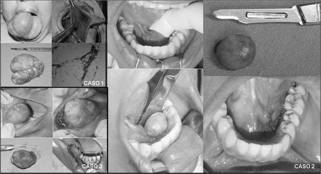
. Case 1: Preoperative, transoperative aspect, macroscopy and histology. Case 2: preoperative, macroscopic aspect of the lesion, transoperative and immediate postoperative aspects. Case 3: Preoperative, transoperative aspect, macroscopy and immediate postoperative aspect.

(0.04MB).
Cases 2 and 3

Cases 2 and 3 are related to smaller lipomas, most commonly found in the oral cavity. In both cases, the patients were females, with ages ranging between 30 and 45 years, respectively. In case 2, the lesion was in the inferior lingual region; and in case 3, it was in the jugal mucosa (Figure 1). Both were treated by surgical excision of the whole lesion and the material was referred to histopathology, with later confirmation of lipoma.

DISCUSSION

Lipomas are histologically very similar to normal fat tissue1. However, its metabolism is quite different from that of the normal tissue, because its lipids are not available for our normal metabolism1. Patients are usually symptom-free, and the lesion is usually a yellowish submucosal mass attached by a sessile base or pedicle5.

It bears variable sizes, from small −10mm masses, to large fat lesions and happen most frequently in the oral mucosa. Tongue, mouth floor, jugal mucosa, vestibule, palate, lips and gums are the most common sites, in descending order5. It more frequently affects persons with more than 40 years of age and bears equal intra-oral gender distribution4.

Although trauma, infection and other factors have been proposed as etiological agents for lipomas, their etiology remains unknown. The treatment of choice is conservative surgical excision and recurrence is rare, as is its malignant transformation4,5.

REFERENCES
[1]
Epivatianos A, Markopoulos AK, Papanayotou P.
Benign Tumors of Adipose Tissue of the Oral Cavity: A Clinicopathologic Study of 13 Cases..
J Oral Maxillofac Surg, 58 (2000), pp. 1113-1117
[2]
Furlong MA, Fanburg-Smith JC, Childers EL.
Lipoma of the oral and maxillofacial region: Site and subclassification of 125 cases..
Oral Surg Oral Med Oral Pathol Oral Radiol Endod, 98 (2004), pp. 441-450
[3]
Fregnani ER, Pires FR, Falzoni R, Lopes MA, Vargas PA.
Lipomas of the oral cavity: clinical findings, histological classification and proliferative activity of 46 cases..
Int J Oral Maxillofac Surg, 32 (2003), pp. 49-53
[4]
Prado R, Ribeiro DPB, Fontoura RA, Sampaio RKPL, Moreira LC.
Lipoma sublingual: relato de caso..
BBO, 55 (1998), pp. 226-228

Paper submitted to the ABORL-CCF SGP (Management Publications System) on March 28th, 2006 and accepted for publication on May 11th, 2006. cod. 1812.

Copyright © 2007. Associação Brasileira de Otorrinolaringologia e Cirurgia Cérvico-Facial
Idiomas
Brazilian Journal of Otorhinolaryngology (English Edition)
Article options
Tools
en pt
Announcement Nota importante
Articles submitted as of May 1, 2022, which are accepted for publication will be subject to a fee (Article Publishing Charge, APC) payment by the author or research funder to cover the costs associated with publication. By submitting the manuscript to this journal, the authors agree to these terms. All manuscripts must be submitted in English.. Os artigos submetidos a partir de 1º de maio de 2022, que forem aceitos para publicação estarão sujeitos a uma taxa (Article Publishing Charge, APC) a ser paga pelo autor para cobrir os custos associados à publicação. Ao submeterem o manuscrito a esta revista, os autores concordam com esses termos. Todos os manuscritos devem ser submetidos em inglês.