
Teratomas are neoplasms derived from germ cells with components of the three embryonic layers (ectoderm, mesoderm, and endoderm), that occur in any age group but are more prevalent in childhood, and have no gender preference.1,2 The lesions can be benign (mature, dermoid, and cystic teratomas) or malignant (immature and solid teratomas), and can affect any structure in the midline).1–3 The clinical presentation varies according to the lesion size and location.1
Imaging studies are useful to show the location and extent of the lesion and to aid in clinical management. Early diagnosis and treatment with excision of the lesion are necessary for a favorable outcome.1,4,5
In the present report, the authors describe a case of nasopharyngeal teratoma, emphasizing diagnosis and treatment-related aspects.
Case reportS.A.R.B., a 1 year 8 month old female infant, was seen with a clinical presentation of nasal obstruction, bilateral purulent nasal discharge, and snoring, all present since she was born. At initial examination, the patient had normal vital signs and exhibited no abnormalities on oropharyngoscopy and otoscopy.
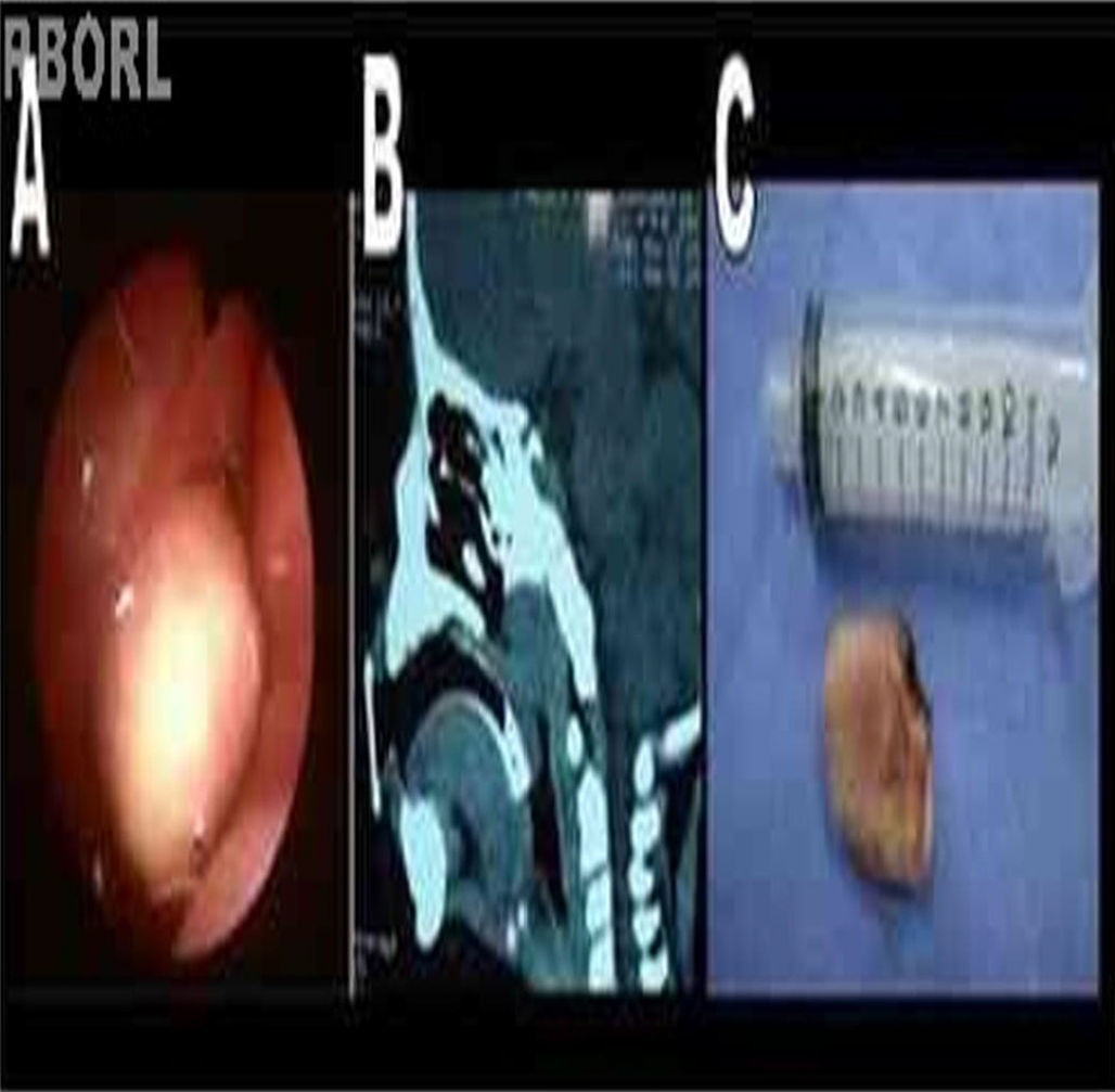
Flexible nasofibroscopy revealed a whitish nasopharyngeal mass (Fig. 1A), completely obstructing the left nasal cavity and partially obstructing the right nasal cavity.
 Tumor obstructing the rhinopharynx on the left. (B) Computed tomography showing a lesion limited to the rhinopharynx. (C) Surgical specimen.")
Computed tomography and magnetic resonance imaging revealed a poorly vascularized obstructive lesion in the nasopharynx, with no signs of infiltration or intracranial extension (Fig. 1B).
Utilizing an endoscopic surgical approach, an electric scalpel was used to uneventfully resect the lesion that arose from the left torus tubarius. The lesion macroscopically appeared similar to a tongue (Fig. 1C). The patient's clinical course improved in the immediate postoperative period.
The histopathological examination identified adipose tissue, mature cartilage tissue, and fibroconnective stroma consisting of skin and skin appendages, forming an epithelial inclusion cyst consistent with a mature teratoma.
DiscussionThis case is relevant both because of its rarity and the importance of the differential diagnosis of nasal obstruction in infants, which should include choanal atresia, intranasal glioma, encephalocele, rhabdomyosarcoma, dermoid cyst, lymphangioma, hemangioma, and neurofibromatosis.
Imaging studies are helpful to determine the differences between solid and cystic tumors, in addition to showing the location and extent of lesions, thus aiding in the clinical management and the surgical approach. However, they do not differentiate benign from malignant lesions.2,3 In the present case, computed tomography (CT) and magnetic resonance imaging (MRI) identified a mass obstructing the nasopharynx, with no signs of infiltration or continuity with intracranial structures.
Knowledge of the limits and size of the tumor are important aspects to be considered in surgical planning.1,2 In the reported case, the surgical excision of the tumor mass was performed endoscopically, with complete resection of the lesion without damage to adjacent structures. The histological examination confirmed the diagnosis of mature teratoma; although the incidence of mature teratomas is 1:4000 live births, it is exceedingly rare in the head and neck and comprises only 2% to 5% of cases.2–5
Final commentsTeratoma must be considered in the differential diagnosis of lesions found in the nasopharynx and nasal cavity, mainly in neonates. Endoscopic and imaging studies (CT and MRI) promote early diagnosis and improve the outcome.
Conflicts of interestThe authors declare no conflicts of interest.
Please cite this article as: Costa CC, Guimarães VD, Moura FS, Chediack MN, Fernandes EJ. Mature teratoma of the nasopharynx. Braz J Otorhinolaryngol. 2014;80:544–5.


