
Allergic rhinitis (AR) is an IgE-mediated non-infectious disease of the nasal mucosa following contact with allergens.
ObjectiveTo investigate the peripheral Th17 cells and CD4+CD25+Foxp3+regulatory T (Treg) cells and the expression of cytokines in the serum of AR patients.
MethodsThe peripheral blood of 14 patients with AR (AR group) and six healthy subjects (control group) was collected from March to May of 2012. Flow cytometry was performed to detect the Th17 cells and Treg cells, and enzyme-linked immunosorbent assay (ELISA) to measure the serum levels of IL-17 and TGF-β1.
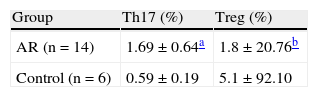
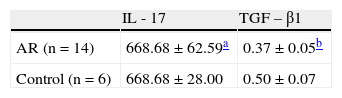
ResultsThe proportion of Th17 cells in the AR group was markedly higher than that in the control group (p<0.01). The proportion of Treg cells in the AR group was also dramatically reduced when compared with the control group (p<0.01). In the AR group, serum IL-17 levels were markedly higher than those in the control group (p<0.01). In the AR group, serum TGF-β1 levels were significantly lower than those in the control group (p<0.01).
ConclusionThe imbalance of peripheral Th17/Treg cells plays an important role in the pathogenesis of AR.
© 2014 Associação Brasileira de Otorrinolaringologia e Cirurgia Cérvico-Facial. Published by ElsevierEditora Ltda. All rights reserved.
A rinite alérgica (RA) é uma doença não infecciosa da mucosa nasal mediada por IgE após o contato com alérgenos.
ObjetivoInvestigar as células Th17 periféricas e CD4+CD25+Foxp3+células T reguladoras (Treg) e a expressão sérica de citocinas em pacientes com RA.
MétodosDe março a maio de 2012, foi coletado o sangue periférico de 14 pacientes com RA (grupo RA) e seis indivíduos saudáveis (grupo controle). A detecção das células Th17 e células Treg foi realizada através da citometria de fluxo e os níveis séricos de IL -17 e TGF- β1. Foram medidos por ELISA.
ResultadosA percentagem de células Th17 no grupo RA foi bem maior do que no grupo controle (p<0,01). A proporção de células Treg no grupo RA também foi drasticamente menor quando comparada ao grupo controle (p<0,01). No grupo RA, o nível sérico de IL-17 foi significativamente maior do que no grupo controle (p<0,01).
ConclusãoO desequilíbrio de células Th17/Treg periféricas desempenha um papel importante na patogênese da RA.
Allergic rhinitis (AR) is an IgE-mediated non-infectious disease of the nasal mucosa following contact with allergens.1 Previous studies have demonstrated that imbalance of Th1/ Th2 cell-mediated immunity has an important role in the pathogenesis of AR, which is characterized by Th2 cell-mediated inflammation.2 In recent years, there is evidence showing that regulatory T (Treg) cells are another subset of T lymphocytes involved in allergic diseases and can regulate the pathological and physiological immune response, which may promote self-immune tolerance and maintenance of immune balance.3 CD4+CD25+ Treg cells are the major type of Treg cells; they can secret some cytokines, including TGF-β1 and IL-10. Forkhead box p3 (Foxp3) gene is a critical regulatory gene in the development and functional maintenance of CD4+CD25+ Treg cells.4
Th17 cells are a subset of T lymphocytes that play crucial roles in pro-inflammatory inflammation and autoimmune diseases. RORγt is a key nuclear transcription factor of Th17 cells, and IL-17 is an important effector cytokine secreted by these cells. IL-17 can exert a pro-inflammatory effect, promote the production of chemokines (such as IL-8 and monocyte chemoattractant protein-1, among others) in the local tissues, facilitate the proliferation of monocytes and neutrophils, and stimulate the generation of IL-6 and prostaglandin E2, resulting in local inflammation.5,6 Some researchers, using real-time polymerase chain reaction (RTPCR) and enzyme-linked immunosorbent assay (ELISA) for the detection of associated transcription factors and cytokines of Th17 and Treg cells in the peripheral blood of AR patients, observed that the expression of RORγt and IL-17 increased and the expression of Foxp3 (transcription factor of Treg cells) and TGF-β1 (associated cytokine) decreased.7 In the present study, flow cytometry (FCM) and ELISA were employed to detect the Th17/Treg cells and serum levels of IL-17 and TGF- β1, respectively, aiming to explore the role of Th17/Treg cells in the pathogenesis of AR.
Materials and methodsSubjectsFrom March to May of 2012, a total 14 patients with AR were admitted into this department: five males and nine females, with a mean age of 29.76years. The diagnosis of AR was made according to its diagnostic criteria.1 Skin- prick test results were positive for dust mite; the severity of AR was moderate or severe in all 14 patients. These patients had not been treated with glucocorticoids in the past month and had not received anti-histamine therapy and allergen-specific immunotherapy. Patients with sinusitis, asthma, and intolerance to acetylsalicylic acid were excluded. Additionally, six healthy volunteers without symptoms of AR were recruited as controls: two males and four females, with a mean age of 30.83years. All subjects signed an informed consent prior to their enrollment in the study.
Main reagents and instrumentFlow cytometer (FACSCalibur; BD - USA) was used for detection and Cellquest software (BD - USA) was employed for the acquisition of data and subsequent analysis. Microplate reader (ELX-800; Bio-Tek - USA), PMA, ionomycin calcium, BFA (MultiSciences), Human Regulatory T Cell Staining Kit (eBioscience - USA), APC-conjugated human CD8 mAb (BD - USA), PerCP-Cy5.5 conjugated human CD3 mAb, PE-conjugated human IL-17 mAb and corresponding istype control (eBioscience - USA), fixation solution and membrane rupture solution (Invitrogen - USA), anti-human IL-17 ELISA kit (eBioscience - USA), and TGF- β1 ELISA kit (RayBio - USA) were used in the present study.
Sample collectionIn the morning, venous blood samples (4mL) were collected from the ulnar vein and anti-coagulated with heparin. Then, 2mL of blood were subjected to centrifugation at 3,000rpm for 15; the serum was collected and stored at -20°C for the detection of IL-17 and TGF-β1. In addition, the remaining 2mL of blood were used for flow cytometry for the detection of Th17 cells and Treg cells.
Detection of Th17 cells by FCMIn brief, 250μL of peripheral blood were mixed with 50μg/L PMA, 2.0μmol/L monensin, and 750 μmoL/L ionomycin calcium, followed by incubation at 37°C in an environment with 50mL/L CO2 for 4h. The cell suspension was transferred into a 1.5ml-PE tube followed by centrifugation at 2,500rpm for 6min. The supernatant was removed, and the cells were washed with PBS twice for FCM. The cells were treated with 10μL of PECy5-CD3Ab and 10μL of FITC-CD8Ab at room temperature, in a dark environment, for 30min.
After washing in PBS twice, the cells were fixed in 300μL of fixation solution in a dark environment at 4°C for 15min. After centrifugation, the supernatant was removed. Following the addition of membrane rupture solution, centrifugation was performed at 3,000rpm and the supernatant was removed. After washing in PBS twice, cells were transferred into two tubes and treated with 20μL of PE-IL-17Ab and 10μL of isotype control PE-IgG1, respectively, in a dark environment at room temperature for 30min. After washing in PBS twice, cells were re-suspended in 0.3mL of PBS and subjected to FCM. CellQuest software was employed for data analysis.
Detection of Treg cells by FCMCells were transferred into sample tube and control tube. Cells in the sample tube were treated with CD4/CD25/ Foxp3, and those in the control group were treated with Foxp3 isotype control (CD4/CD25/mouse IgG). Then, 100μL of anti-coagulated blood was added followed by incubation in a dark environment at room temperature for 20min. Following the addition of 1mL of hemolytic agent, incubation was performed at room temperature in a dark environment for 10min followed by centrifugation at 1,000rpm for 5min. The supernatant was removed and cells were re-suspended in 1mL of PBS followed by centrifugation at 1,000rpm for 5min. The supernatant was then removed, and 0.5mL of Foxp3 fixation solution were added, followed by incubation in a dark environment at room temperature for 20min. Cells were re-suspended in 1mL of PBS, and centrifuged at 1,000rpm for 5min. The supernatant was removed, and 0.5mL of Foxp3 membrane rupture solution was added to each tube, followed by centrifugation at 1,000rpm for 5min. These cells were re-suspended in 0.5mL of Foxp3 membrane rupture solution, followed by incubation in a dark environment at room temperature for 15min. After centrifugation at 1,000rpm for 5min, the supernatant was removed. Then, 10μL of PE Foxp3Ab were added to the sample tube and 10μL of isotype control were added to the control tube, followed by incubation at room temperature in a dark environment for 30min. Cells were re-suspended in 1mL of PBS and centrifuged at 1,000rpm for 5min. The supernatant was removed and the cells were re-suspended in 0.4mL of PBS. FCM was performed and CellQuest software was employed for data analysis.
Detection of serum IL-17 and TGF- β1 by ELISAThe detection of serum contents of IL-17 and TGF- β1 was performed according to the manufacturer’s instructions. The lower limit of detection was 0.5pg/mL for IL-17 and 9pg/mL for TGF- β1.
Statistical analysisStatistical analysis was performed with SPSS version 16.0. Data with normal distribution were expressed as mean±standard deviation. Student’s t-test for independent variables was performed for comparisons between two groups. A value of p<0.05 was considered statistically significant.
ResultsProportion of Th17 cells in AR group and control groupFCM demonstrated that the proportion of Th17 cells in the AR group was markedly higher than that of the control group (p<0.01; Table 1).
Proportion of Treg cells in AR group and control groupIn the AR group, the proportion of Treg cells was markedly reduced when compared to the control group (p<0.01; Table 1).
Serum contents of IL-17 and TGFpi in AR group and control groupELISA was performed to detect the serum contents of IL-17 and TGF- β1 in the AR group and control group. Data are shown in Table 2. Results demonstrated that the IL-17 level in the AR group was markedly higher than that of the control group (p<0.01). In the AR group, the TGF- β1 level was dramatically reduced when compared with the control group (p<0.01).
DiscussionPrevious studies have demonstrated that the serum level of IL-17 in AR patients was significantly increased when compared with controls,8 and that the proportion of IL-17 positive cells in the nasal mucosa in AR patients was also markedly higher than that in healthy subjects.9 Ciprandi et al.10,11 observed that the serum IL-17 level was positively related to the severity of symptoms of AR and positively associated with the number of peripheral eosinophils. The peripheral monocytes from AR patients who were treated with pollen allergen and FCM showed the proportion of Th17 cells was markedly higher than that in the control group. In the present study, the results indicated that the proportion of peripheral Th17 cells in AR patients was significantly higher than that of the controls, accompanied by the increase in serum level of IL-17. These findings suggest that Th17 cells can secret a large amount of IL-17, which is involved in the pathogenesis of AR.
Additionally, the proportion of CD4+CD25+Treg cells in peripheral blood of AR patients was significantly lower than that of the controls, accompanied by the reduction of serum TGF- β1 level. This suggests that the reduction of peripheral Treg cell count leads to a compromised function of these cells. In healthy subjects, the proportion of CD4+CD25+Treg cells was approximately 5% to 10% in the peripheral blood. The CD4+CD25+Treg cells can exert inhibitory effect on the immune response via the intercellular interaction or secretion of inhibitory cytokines (such as IL- 10 and TGF-β).4 The present results demonstrated that the proportion of peripheral CD4+CD25+Treg cells and the serum level of TGF- β1 were reduced. Xu et al.12 confirmed that the Foxp3+positive lymphocytes and Foxp3 mRNA expression in the nasal mucosa and peripheral monocytes of AR patients were markedly reduced when compared with healthy controls, and that Foxp3 and CD4+CD25+Treg cells in allergy patients were markedly reduced.13 This suggests that the compromised inhibitory effect of Treg cells leads to the occurrence of allergic diseases.
Th17 cells and Treg cells are derived from naïve T cells. Th17 cells mediate the inflammatory reaction, whereas Treg cells mediate immune tolerance. There is antagonism in the function and differentiation of Th17 cells and Treg cells. Thus, the balance of Treg cells and Th17 cells is crucial for the maintenance of the immune status. In the present study, the results demonstrated that the proportion of Th17 cells in the peripheral blood of AR patients was markedly increased and that of Treg cells was dramatically reduced. This suggests that Th17/Treg imbalance plays an important role in the pathogenesis of AR. Further research in the Th17/ Treg balance may provide a novel strategy for the treatment of AR.
Conflicts of interestThe authors declare no conflicts of interest.
Please cite this article as: Huang X, Chen Y, Zhang F, Yang Q, Zhang G. Peripheral Th17/Treg cell-mediated immunity imbalance in allergic rhinitis patients. Braz J Otorhinolaryngol. 2014;80:152-5
Study conducted at Third Hospital, Sun Yat-sen University, Guangdong, China
gology is pleased to honor the reviewers


