
The cysts that originate in the remnants of dental lamina or enamel organ are known as odontogenic cysts. They are of great interest to otolaryngologists due to their proximity to the maxillary sinus and adjacent areas.1 Radicular cysts represent 72.5% of all odontogenic cysts, followed by dentigerous cysts (DC) (22.2%), and residual cysts (RC) (4.26%).2 The options for treatment of cystic lesions involve curettage, enucleation, marginal resection,3 and endoscopic surgery, which is a viable alternative, especially in cases of extension to the maxillary sinus.4 A PubMed and EBSCOhost search did not retrieve any studies analyzing the simultaneous occurrence of DC and RC. This article reports a case involving the association a DC and a RC in the upper jaw of an edentulous patient.
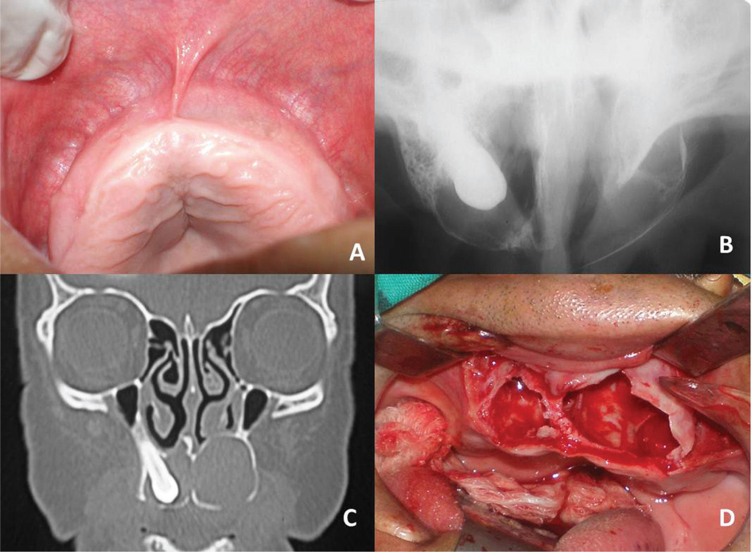
Case presentationA 73-year-old man, asymptomatic, was referred to this clinic for evaluation of an intra-oral swelling. During the extra-oral examination, no volumetric changes or asymmetries were observed. The oroscopy evidenced a discrete swelling in the left buccal area in comparison with the right side, and it had a crackling consistency. The mucosa was normal, but a purplish color suggestive of intraosseous lesion was observed. In the palatal region, the swelling was bilateral, and it also had a crackling consistency (Fig. 1A). In the occlusal radiography, bilateral radiolucent areas with expansion of the buccal cortical were observed on the left side, and a canine tooth was found on the right side. (Fig. 1B). In the computed tomography (coronal view), two hypodense lesions were observed, separated by a hyperdense thin bone wall. The involvement of the canine tooth in the lesion on the right side was clear. The nasal fossa floor on the left side presented as elevated (Fig. 1C). Based on these findings, the primary diagnosis was DC on the right side and RC on the left side. Under general anesthesia, the lesions with enucleation were treated (Fig. 1D), and a primary closure of the wounds was performed. The microscopic examination confirmed the diagnosis of DC and RC.

A, Palatal bilateral swelling. B, Radiolucent area with expansion of the buccal cortical on the left side, and a canine tooth associated with a radiolucent area on the right side. C, Hypodense areas in the coronal view of computed tomography. An elevation in the left nasal fossa floor is visible. D, Surgical cavities.
No reports of an association between DC and RC were retrieved on a PubMed and EBSCOhost search. The cysts are often asymptomatic, and are found on routine examination. Their growth promotes bone resorption, leaving the bone that covers them very thin, as reported in the present study. DC are more frequent in young patients,2 and the rarity of the association between DC and RC is probably a result of the unusual diagnosis of DC in elderly patients. The development of cystic lesions may lead to bone resorption, dental damage, and harm to the anatomical structures (maxillary sinus, nasal cavity, and mandibular fractures), especially when they reach large proportions. The neoplastic transformation of the epithelial lining of odontogenic cysts is rare, although well-described in the literature.5 The presence of an RC in an edentulous area prevents the functional rehabilitation of the patient due to the resulting bone defect.6 All these factors justify the need for the surgical treatment of odontogenic cysts. In the present study, the lesion reached great proportions, which suggests a long course of evolution, despite the fact that the patient was asymptomatic.
Final remarksOdontogenic cysts are commonly observed in jaws, usually causing damage to adjacent structures. Even though they may reach great proportions, they do not always cause pain, which highlights the importance of a thorough clinical examination even in edentulous areas. The association between DC and RC has not been previously described, and in this paper, the authors warned of the possibility and present the surgical treatment of the lesions.
Conflicts of interestThe authors declare no conflicts of interest.
Please cite this article as: Nogueira AS, Sampieri MBS, Sanches-Gonçales ES, Barreto-Gonçales Andrea Guedes, Soares ECS. Simultaneous occurrence of dentigerous cyst and residual cyst in the maxilla. Braz J Otorhinolaryngol. 2014;80:88–9.


