
Binder syndrome is a rare congenital malformation (maxillonasal dysplasia), which was first described in 1939 by Noyes, whereas it was Binder who defined it as a syndrome in 1962.1,2 Clinically, there is enlarged nasal angle (arhinoid face), abnormal position of the nasal bones, hypoplastic maxillary, hypoplasia/reduction of the nasal spine, absence/hypoplasia of the frontal nasal sinus (not obligatory), and atrophy of the nasal mucosa.2,3 In addition, some cases (40–50%) may present malformation in cervical vertebrae – most frequently, the C1 and C2 vertebrae are affected.3
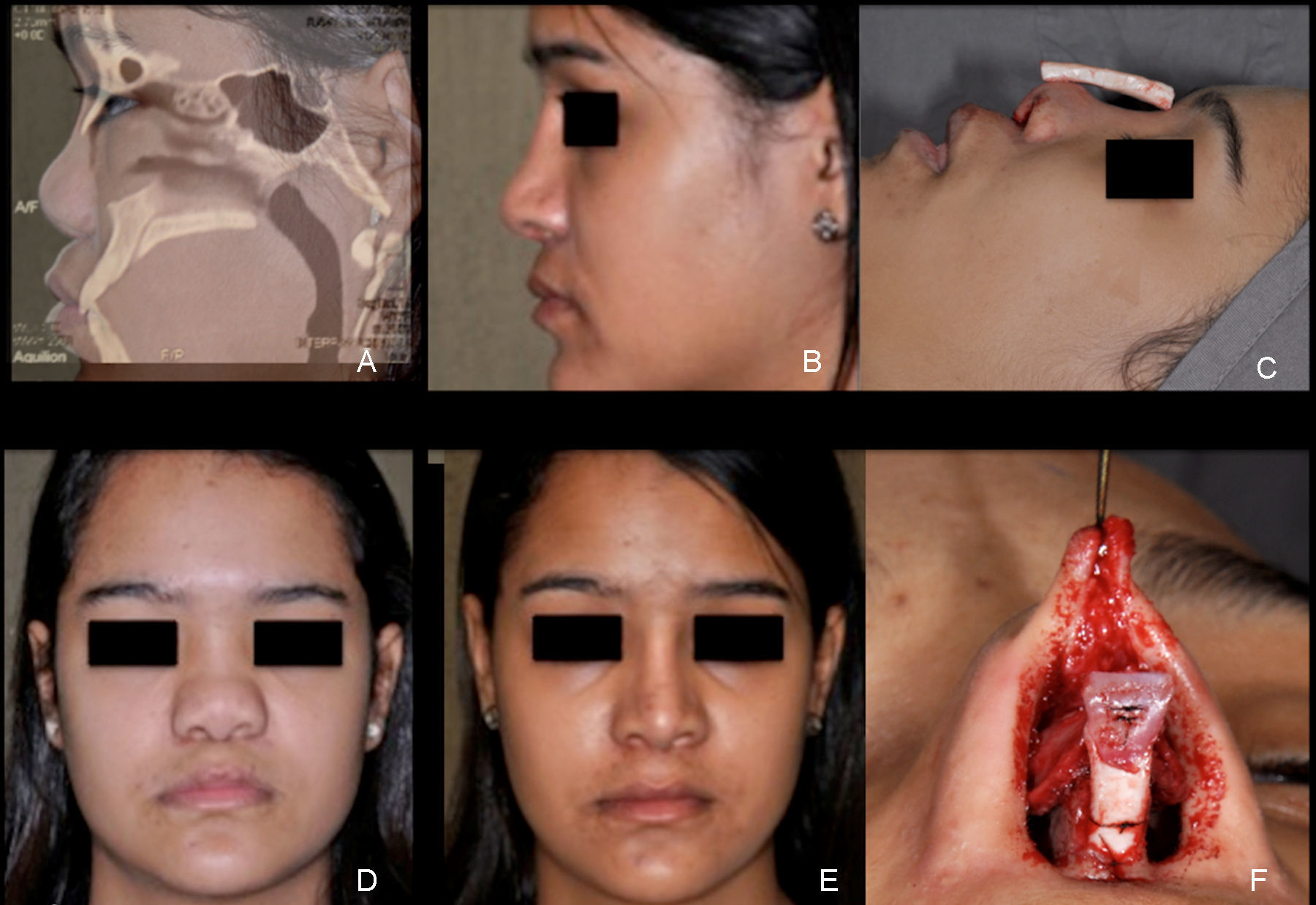
Case reportA 16 year-old female patient came to our department with a complaint of “midfacial retrusion and flat nose” since childhood. She denied nasal obstruction, trauma, previous surgery and comorbidities. Physical examinations revealed the presence of convex lip, nasomaxillary hypoplasia, plan nasofrontal angle, acute nasolabial angle, nasal mucosa atrophy, triangular nostrils and flat nose (Fig. 1A and D). A computer tomography of the paranasal sinuses was performed and revealed absence of nasal spine, reduced horizontal dimensions of the jaw, nasal septal cartilage aplasia, obtuse nasofrontal angle, and retracted position of the jaw relative to the cranial base (Fig. 1A). No cervical disease was found.
 Preoperative profile and sagittal CT. (B) Postoperative profile view. (C) Intraoperative: costal cartilage for onlay dorsum graft. (D) Preoperative frontal view. (E) Postoperative frontal view. (F) Intraoperative: tip and columellar grafts.")
The diagnosis of Binder syndrome was made according to clinical and radiological findings. Augmentation rhinoplasty was performed with autologous costal cartilage and remaining septal cartilage. The goal was to increase the nasal dorsum, to allow tip projection and support, projection of the premaxillary region and increase nasal lengthening. The following grafts were carved: dorsal onlay graft with costal cartilage, extended columelar strut, lateral crural strut graft, Sheen Shield graft and premaxillary graft (Fig. 1C and F). The two-month postoperative result can be seen in Fig. 1B and E, with adequate gain in dorsum projection, tip and premaxilla. The esthetic and functional results were quite satisfactory.
DiscussionBinder syndrome is a rare congenital deformity, and the most common characteristics presented are flattened nose and midfacial retrusion, as we saw on the patient described.
The diagnosis of Binder syndrome is made on the basis of standard clinical and radiologic findings. Genetic review can also be helpful.4 The real cause of this disease is still obscure, though the inhibition of the ossification center that would normally form the lateral and inferior borders of the piriform aperture during the fifth and sixth week of pregnancy, leading to a localized hypoplasia of the upper jaw and thus resulting in a retracted columellar/lip junction and lack of the normal triangular flare in the lower part of the columella, has been suggested as cause.4 However, the etiology of the syndrome is not yet clear; it is believed that it is an association of both the genetic and environmental factors (vitamin K deficiency during pregnancy). Birth trauma has also been proposed.
The treatment is controversial and should be performed according to the age and disease severity. Patients with milder forms of the disease may benefit from rhinoplasty only, as the case presented here. Moreover, the Class 3 malocclusion patients require orthognathic correction before nasal reconstruction.5
ConclusionThe case report presents the craniofacial characteristics compatible with Binder syndrome, as supported by the literature. The knowledge of ideal proportions of the face helped us to achieve the correct diagnosis of the syndrome and its proper treatment.
Conflicts of interestThe authors declare no conflicts of interest.
Please cite this article as: Arroyo HH, Olivetti IP, Santos VG, Weber R, Jurado JR. Nasal reconstruction in Binder syndrome. Braz J Otorhinolaryngol. 2017;83:488–9.
Peer Review under the responsibility of Associaçao Brasileira de Otorrinolaringologia e Cirurgia Cervico-Facial.
gology is pleased to honor the reviewers


