




Schwannomas are benign peripheral nerve sheath tumors that infrequently arise in the head and neck, with involvement of the superior laryngeal nerve (SLN) being exceedingly rare.1,2 The external branch of the SLN (EBSLN) innervates the cricothyroid muscle, which is essential for vocal fold tension and pitch control. Consequently, injury to this nerve can result in subtle dysphonia and an increased risk of aspiration. Preoperative diagnosis remains challenging, as EBSLN lesions lie deep within the carotid space and closely mimic other neurogenic or vascular neoplasms. Herein, we report a rare case of an EBSLN schwannoma managed by en bloc excision requiring complete nerve transection. This was immediately followed by primary end-to-end neurorrhaphy, and we emphasize the favorable short-term functional outcomes achieved through this reconstructive strategy.
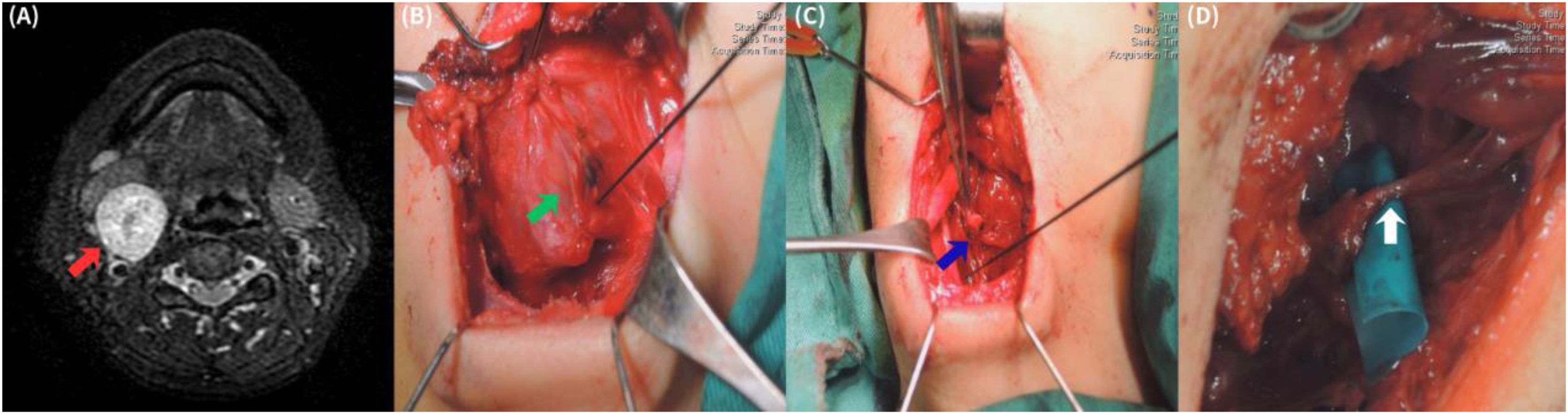
Case reportA 50-year-old woman with no major comorbidities presented with a slowly enlarging, mildly tender right level II neck mass of a 2-year duration. Physical examination revealed a mobile mass measuring approximately 2 × 2 cm. Magnetic resonance imaging (MRI) demonstrated a 3.5 × 3.1 × 2.9 cm carotid-space lesion suggestive of a neurogenic tumor (Fig. 1A). Preoperative laryngoscopy revealed normal bilateral vocal fold mobility without any preexisting phonatory disability.

Standard intermittent intraoperative neuromonitoring (I-IONM) was utilized during the procedure to map the nerve, facilitate safe tumor dissection, and verify the functional integrity of the neural pathways before and after resection. The tumor was identified as arising from the external branch of the right superior laryngeal nerve (EBSLN) (Fig. 1B). Intraoperatively, the EBSLN was identified as Type 1 according to Cernea's classification, crossing the superior thyroid artery more than 1 cm above the upper pole of the thyroid gland.3 To achieve en bloc excision of the tumor, a complete nerve transection was performed (Fig. 1C). Immediate tension-free, end-to-end microsurgical neurorrhaphy was then performed to reconstruct the neural defect (Fig. 1D). Histopathology revealed a well-defined nodular lesion featuring both Antoni A and B areas with Verocay bodies. Immunohistochemical staining was positive for S100 and SOX10, confirming a completely excised benign schwannoma. Postoperatively, a liquid diet was tried on day 1, and a regular diet was smoothly resumed subsequently. The drainage tube was removed on postoperative day 5, and the patient was discharged without any clinical signs of aspiration, dysphagia, or other complications.
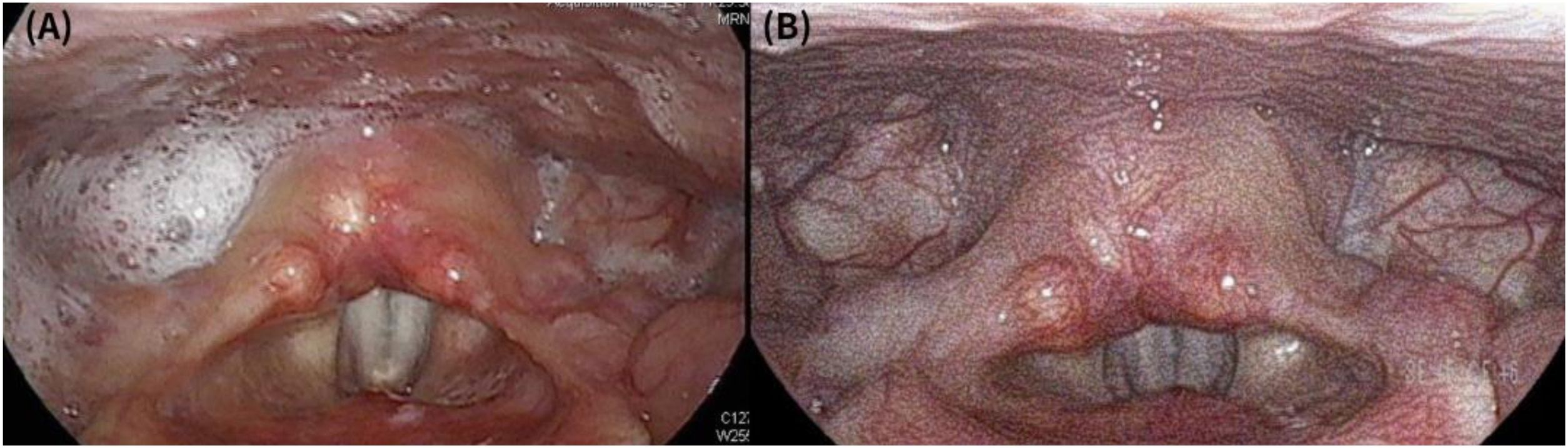
At the 1-month follow-up, laryngoscopy revealed intact gross vocal fold mobility but notable saliva pooling in the right pyriform sinus (Fig. 2A). At the 2-month follow-up, formal speech evaluation quantified the mild phonatory difficulty. The perceptual evaluation yielded a GRBAS score of 1 (G1 R0 B1 A1 S1), indicating mild hoarseness, breathiness, and vocal strain. Objective acoustic analysis recorded a fundamental frequency (F0) of 202.6 Hz and a maximum phonation time of 11.4 seconds. At the 3-month follow-up, laryngoscopy demonstrated complete resolution of the saliva pooling and optimal vocal fold tension, with marked subjective improvement in both phonation and swallowing (Fig. 2B).
Discussion
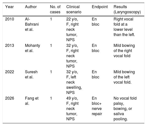
Schwannomas of the superior laryngeal nerve (SLN) are exceedingly uncommon, and isolated involvement of its external branch (EBSLN) is even rarer, often presenting diagnostic and therapeutic challenges.1,2 As highlighted initially, while the precise anatomical origin from the EBSLN is established through preoperative MRI and intraoperative findings, the definitive histopathological diagnosis strictly relies on microscopic examination and immunohistochemistry. A summary of previously selected reported cases highlights both the extreme rarity of this condition and the prevailing surgical management trends (Table 1).
Summary of selected reported cases of external branch of the superior laryngeal nerve (EBSLN) schwannomas.
| Year | Author | No. of cases | Clinical scenario | Endpoint | Results (Laryngoscopy) |
|---|---|---|---|---|---|
| 2010 | Al-Bahrani et al. | 1 | 22 y/o, F, right neck tumor, NPS | En bloc | Right vocal fold at a lower level than the left. |
| 2013 | Mohanty et al. | 1 | 32 y/o, F, right neck tumor, NPS | En bloc | Mild bowing of the right vocal fold |
| 2022 | Suresh et al. | 1 | 32 y/o, F, left neck swelling, NPS | En bloc | Mild bowing of the left vocal fold. |
| 2026 | Fang et al. | 1 | 49 y/o, F, right neck tumor, NPS | En bloc+ nerve repair | No vocal fold palsy, bowing, or saliva pooling. |
Abbreviations: F, Female; NPS, No Phonatory Symptoms.
The fundamental challenge in managing these carotid space tumors lies in the EBSLN’s critical physiological role. The nerve primarily innervates the cricothyroid muscle, which is essential for vocal fold tension and pitch modulation.4 Even subtle injury during tumor extirpation can result in vocal fatigue, restricted pitch range, and mild aspiration. Consequently, when complete tumor clearance inevitably requires complete nerve transection, surgeons face a critical dilemma between adequate excision and functional preservation.
To resolve this dilemma, we advocate the routine use of intraoperative nerve monitoring (IONM) coupled with immediate neural reconstruction. In our case, IONM was pivotal for confirming the functional integrity of the proximal and distal nerve stumps prior to tension-free, epineurial end-to-end neurorrhaphy.5 A unique postoperative observation following the nerve sacrifice was transient saliva pooling in the pyriform sinus. While supraglottic sensation is primarily mediated by the internal SLN, we hypothesize that temporary denervation of the cricothyroid muscle causes a subtle loss of anterior laryngeal suspension, altering pharyngolaryngeal coordination and decreasing the efficiency of pyriform sinus clearance. The complete resolution of pooling by three months strongly correlates with the timely reinnervation of the cricothyroid muscle facilitated by our immediate repair.
Historically, reports of SLN tumor resection frequently described postoperative vocal bowing and dysphagia with highly variable recovery trajectories, primarily because reconstruction was rarely attempted.1,2 However, recent literature demonstrates that immediate anastomosis for EBSLN or recurrent laryngeal nerve defects significantly improves vocal outcomes and prevents irreversible muscle atrophy.6 By minimizing the denervation interval, primary neurorrhaphy—or interposition grafting (e.g., ansa cervicalis) when direct repair is unfeasible—preserves the motor endplates of the cricothyroid muscle.4 Our sequential laryngoscopic evidence validates that early surgical reconstruction, aligned with targeted rehabilitation, is a highly effective strategy to optimize functional recovery.
ConclusionComplete surgical excision of an external branch of the superior laryngeal nerve (EBSLN) schwannoma often necessitates nerve sacrifice, posing a significant risk to vocal and swallowing functions. This case demonstrates that immediate, tension-free end-to-end neurorrhaphy, guided by intraoperative nerve monitoring (IONM), is a highly effective reconstructive strategy. By minimizing the denervation interval, this proactive approach successfully preserves cricothyroid muscle innervation and prevents long-term pharyngolaryngeal dysmotility, offering patients an optimal trajectory for functional recovery.
ORCID IDKo-Chun Fang: 0000-0003-4735-0799
Chi-Chih Lai: 0000-0002-5281-2911
Authors’ contributionsKo-Chun Fang: data collection, manuscript drafting, and figure preparation; Chi-Chih Lai: conceptualization, supervision, and manuscript revision. All authors have read and approved the final version to be published and agree to be accountable for all aspects of the work.
Patient consent and ethicsWritten informed consent for publication was obtained from the patient. This study was approved by the Chang Gung Medical Foundation.
Institutional Review Board (Ethical Application Reference number: 202500528B0).
Declaration of Generative AI and AI-assisted technologies in the writing processDuring the preparation of this manuscript, the authors used ChatGPT (OpenAI) to assist with English language editing and improvement of clarity.
FundingNone.
Data availabilityThe authors declare that all data are available in repository.
The authors declare no conflicts of interest.




