





The causal relationship between Autoimmune Diseases (ADs) and Hearing Loss (HL) remains unclear. This study investigates whether genetic predispositions associated with ADs contribute to HL risk.
MethodsMendelian Randomization (MR) analysis was conducted to explore the causal effects of ADs on HL. SNPs from Genome-Wide Association Studies (GWAS) were used as instrumental variables for ADs, including Rheumatoid Arthritis (RA), Type 1 Diabetes (T1D), Systemic Lupus Erythematosus (SLE), Sjögren’s Syndrome (SS), Ankylosing Spondylitis (AS), Multiple Sclerosis (MS), Crohn's Disease (CD), and Ulcerative Colitis (UC). Outcome data included Sensorineural Hearing Loss (SNHL), Conductive Hearing Loss (CHL), Mixed conductive and sensorineural Hearing Loss (MHL), and Sudden Idiopathic Hearing Loss (SIHL). MR analyses employed Inverse Variance Weighted (IVW) as the primary method, supplemented with MR-Egger, weighted median, and weighted mode. Heterogeneity, pleiotropy, and sensitivity were evaluated using Cochran's Q test, MR-Egger regression, MR-PRESSO, and leave-one-out analysis.
ResultsThe IVW method identified nine significant associations: MS-SIHL (OR = 1.0494, 95% CI 1.0072–1.0934), AS-CHL (OR = 1.2832, 95% CI 1.0643–1.5472), AS-MHL (OR = 1.5994, 95% CI 1.3696–1.8678), AS-SNHL (OR = 1.1903, 95% CI 1.1104–1.276), AS-SIHL (OR = 1.481, 95% CI 1.22–1.798), SLE-CHL (OR = 1.0593, 95% CI 1.0116–1.1092), UC-MHL (OR = 1.0907, 95% CI 1.0027–1.1865), CD-CHL (OR = 1.0529, 95%CI: 1.0074–1.1005), and CD-SIHL (OR = 1.0597, 95% CI 1.0177–1.1034). Among these, outliers were detected only in AS-SNHL. After outlier removal, the AS-SNHL association remained significant (OR = 1.1722, p < 0.00001), with resolved heterogeneity and pleiotropy. No heterogeneity and pleiotropy were found for the other associations.
ConclusionThis study identified nine significant AD-HL associations, emphasizing the need for targeted screening and management of HL in individuals with AD.
Level of EvidenceLevel 5.
Hearing Loss (HL) is a common sensory disorder affecting individuals of all ages, with significant impacts on quality of life. It is broadly classified into Sensorineural Hearing Loss (SNHL), Conductive Hearing Loss (CHL), and Mixed conductive and sensorineural Hearing Loss (MHL).1 SNHL, caused by damage to the cochlea or auditory nerve, is the most common type, with sudden cases (Sudden Idiopathic Hearing Loss; SIHL) affecting 5–27 per 100,000 annually in the United States and prevalence increasing significantly with age.2 CHL, caused by impaired sound transmission in the outer or middle ear, commonly arises from otitis media and affects approximately 1.7% of children globally, with a substantially higher burden in low- and low-middle-income regions.3 MHL combines elements of both. Risk factors for HL include genetic predisposition, infections, ototoxic medications, noise exposure, and systemic conditions such as Autoimmune Diseases (ADs).4,5
ADs are conditions characterized by immune system dysregulation, leading to attacks on the body’s own tissues, with examples including Rheumatoid Arthritis (RA), Type 1 Diabetes (T1D), Systemic Lupus Erythematosus (SLE), and Sjögren’s Syndrome (SS).6 These diseases share mechanisms like autoantibody production, chronic inflammation, and cytokine imbalances, resulting in tissue damage and impaired immune tolerance.7 ADs are increasingly recognized as risk factors for HL due to their potential to mediate immune-driven damage to auditory structures.8 Mechanistically, autoantibodies and inflammatory cytokines disrupt the blood-labyrinth barrier, impair cochlear microcirculation, and induce oxidative stress, leading to hair cell apoptosis and auditory dysfunction.9 Chronic inflammation further destabilizes neural signaling and endolymphatic homeostasis, exacerbating HL.10 These processes highlight the vulnerability of auditory structures to immune-mediated damage.
While the association between ADs and HL has been reported, the causal relationship remains unclear. To address this, Mendelian Randomization (MR) analysis offers a robust approach to investigate potential causality between ADs and HL. MR utilizes genetic variants, typically Single Nucleotide Polymorphisms (SNPs) identified through Genome-Wide Association Studies (GWAS), as Instrumental Variables (IVs) to assess the causal relationship between an exposure and an outcome.11 By minimizing confounding and reverse causality, MR provides a reliable method to identify causal links that may not be feasible to explore through randomized controlled trials.12 This study employs a two-sample MR approach to investigate the genetically predicted causal relationship between ADs and HL. The findings may contribute to a better understanding of their connection and inform future research on preventive and therapeutic approaches.
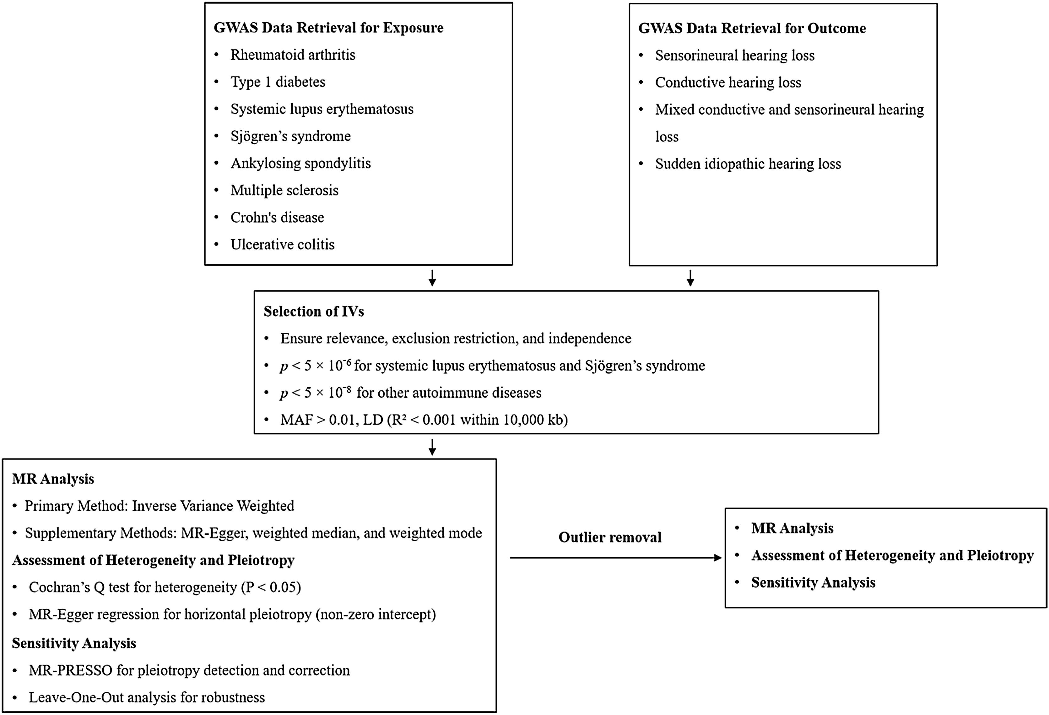
MethodsStudy designThe methodology followed the STROBE-MR statement guidelines. Relevant GWAS databases were selected to obtain the appropriate SNPs. IVs were selected based on the three key principles of MR analysis: relevance, independence, and exclusion restriction.13 MR analysis was then performed to investigate causal relationships. Sensitivity was assessed using various statistical methods to ensure the robustness of the findings, providing a thorough understanding of the potential associations between ADs and HL risk. The study flowchart is illustrated in Fig. 1.

Study flowchart for Mendelian Randomization (MR) analysis of Autoimmune Diseases (ADs) and Hearing Loss (HL). The flowchart outlines the methodology for investigating causal relationships between ADs and various types of HL using MR. Genome-Wide Association Study (GWAS) data retrieval included datasets for Rheumatoid Arthritis (RA), Type 1 Diabetes (T1D), Systemic Lupus Erythematosus (SLE), Sjögren's Syndrome (SS), Ankylosing Spondylitis (AS), Multiple Sclerosis (MS), Crohn’s Disease (CD), and Ulcerative Colitis (UC), as well as outcomes for Sensorineural Hearing Loss (SNHL), Conductive Hearing Loss (CHL), Mixed conductive and sensorineural Hearing Loss (MHL), and Sudden Idiopathic Hearing Loss (SIHL). Instrumental Variable (IV) selection ensured relevance, independence, and exclusion restriction, with strict thresholds for Single Nucleotide Polymorphism (SNP) significance. MR analysis primarily used the Inverse Variance Weighted (IVW) method, supplemented by MR-Egger, weighted median, and weighted mode. Sensitivity analyses included Mendelian Randomization Pleiotropy Residual Sum and Outlier (MR-PRESSO) for pleiotropy correction, leave-one-out analysis for robustness, and heterogeneity tests using Cochran’s Q and MR-Egger regression. Outlier removal was followed by a second round of MR analysis, along with reassessment of heterogeneity, pleiotropy, and sensitivity.
Exposure data were sourced from the IEU Open GWAS database and included eight common ADs: RA, T1D, SLE, SS, Ankylosing Spondylitis (AS), Multiple Sclerosis (MS), Crohn's Disease (CD), and Ulcerative Colitis (UC) based on a published study.14 Outcome data on HL were derived from the FinnGen R11 database (https://www.finngen.fi/en/access_results), including SNHL, CHL, MHL, and SIHL. Both exposure and outcome GWAS were restricted to individuals of European ancestry to minimize population stratification. Outcome data were obtained from Finnish participants, whereas exposure data were not country-specific. All GWAS data used in this study were derived from previously published studies with documented ethical approval and informed consent. Detailed information on these datasets is provided in Table S1.
IV selectionSNPs significantly associated with SLS and SS were included with a threshold of p <5 × 10⁻⁶, due to difficulties in identifying sufficient SNPs that meet stricter significance levels. For other ADs, a threshold of p <5 × 10⁻⁸ was applied.15 Only SNPs with a Minor Allele Frequency (MAF) >0.01 were retained.16 Linkage Disequilibrium (LD) was minimized by excluding SNPs with R2 > 0.001 within a 10,000 kb window.17 Proxy SNPs with R2 > 0.8 were used if IVs were absent in the outcome data.18 F-statistics (F = R2 × (N-2) / (1-R2)) were calculated to ensure IV strength (F > 10) and prevent weak instrument bias. R² represents the proportion of variance in the exposure explained by the SNP.19
MR analysisThe Inverse Variance Weighted (IVW) random effects method was the primary approach, providing Odds Ratios (ORs) with 95% Confidence Intervals (CIs) by calculating a weighted average effect size based on SNP inverse variance.20 To ensure robustness, MR-Egger, weighted median, and weighted mode methods were applied. MR-Egger accounts for pleiotropy by including an intercept term,21 the weighted median provides reliable estimates if at least 50% of IVs are valid,22 and the weighted mode identifies the causal effect as the precision-weighted mode, even when most IVs are invalid.23
Sensitivity analysisSensitivity analyses were performed to ensure the robustness of the findings and detect pleiotropy. Heterogeneity was assessed using the IVW method with Cochran's Q test. A p-value >0.05 indicates low heterogeneity, suggesting a minimal influence on IVW results.24 Pleiotropy was evaluated using MR-Egger regression and the MR-PRESSO. MR-Egger regression assessed horizontal pleiotropy through its intercept, with values close to zero or non-significant indicating no pleiotropy.25 MR-PRESSO identified and corrected horizontal pleiotropy by excluding outlier SNPs.26 Leave-one-out analysis was performed by sequentially removing each SNP to evaluate its influence on the overall estimates and assess the stability of the results.27
Software and data visualizationForest, scatter, and funnel plots were used for visualization. All analyses were performed using R version 4.0.5 and the “TwoSampleMR” package.
ResultsIV selectionThe study selected 21 IVs for UC, 36 IVs for T1D, 15 IVs for SLE, 10 IVs for SS, 25 IVs for RA, 22 IVs for MS, 52 IVs for CD, and 26 IVs for AS. The mean F-values for these IVs ranged from 30.491 for SLE to 153.719 for AS, indicating strong instrument strength (Table S2). Proxy SNPs were identified for SIHL, SNHL, MHL, and CHL across various exposures. For example, rs2853953 replaced rs1131114 in SLE for all four outcomes (Table S3). Palindromic SNPs were identified for various exposures, including rs12692254 for CD, rs3093017 for RA, and rs4065985 and rs7936434 for UC (Table S4).
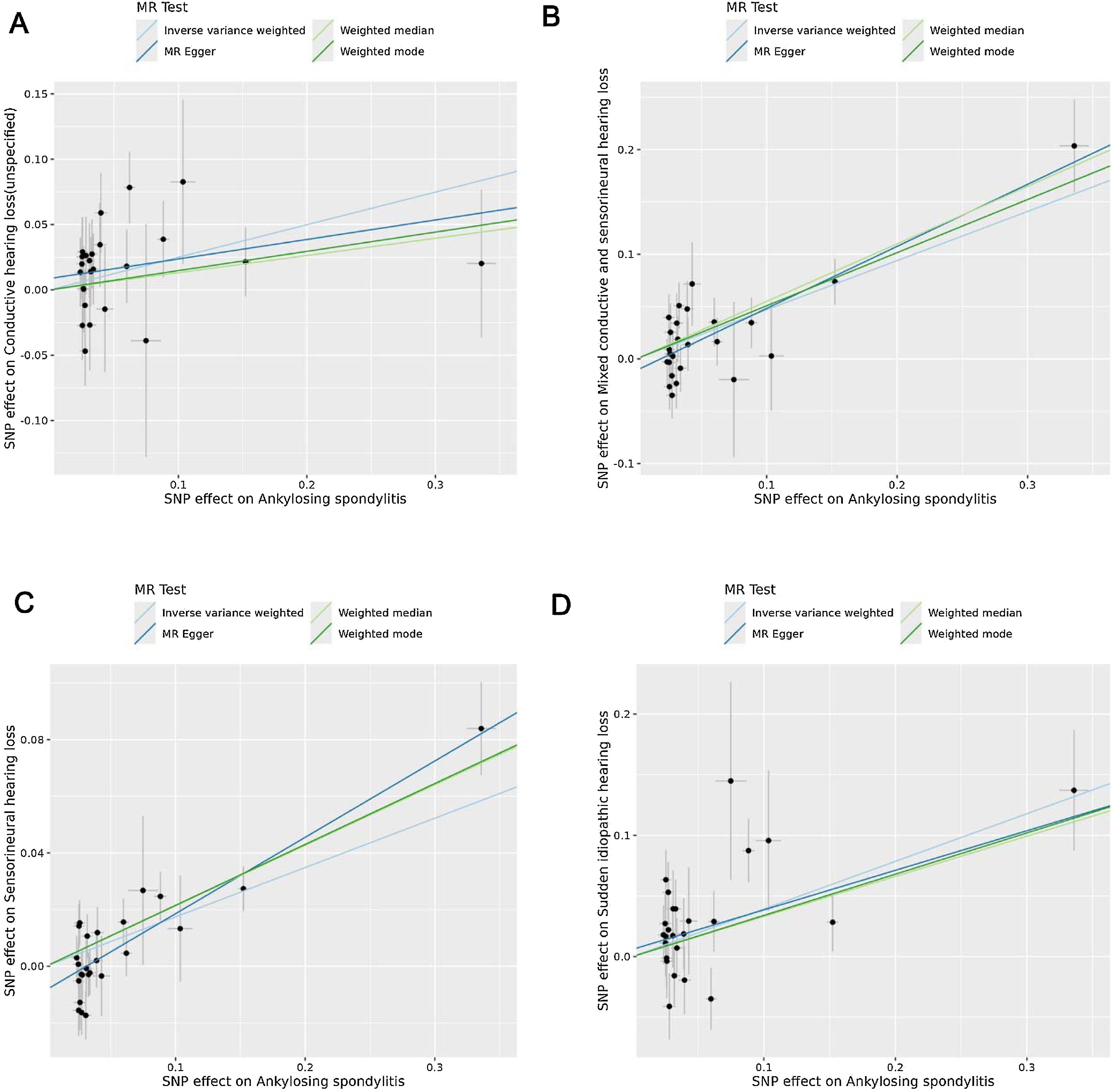
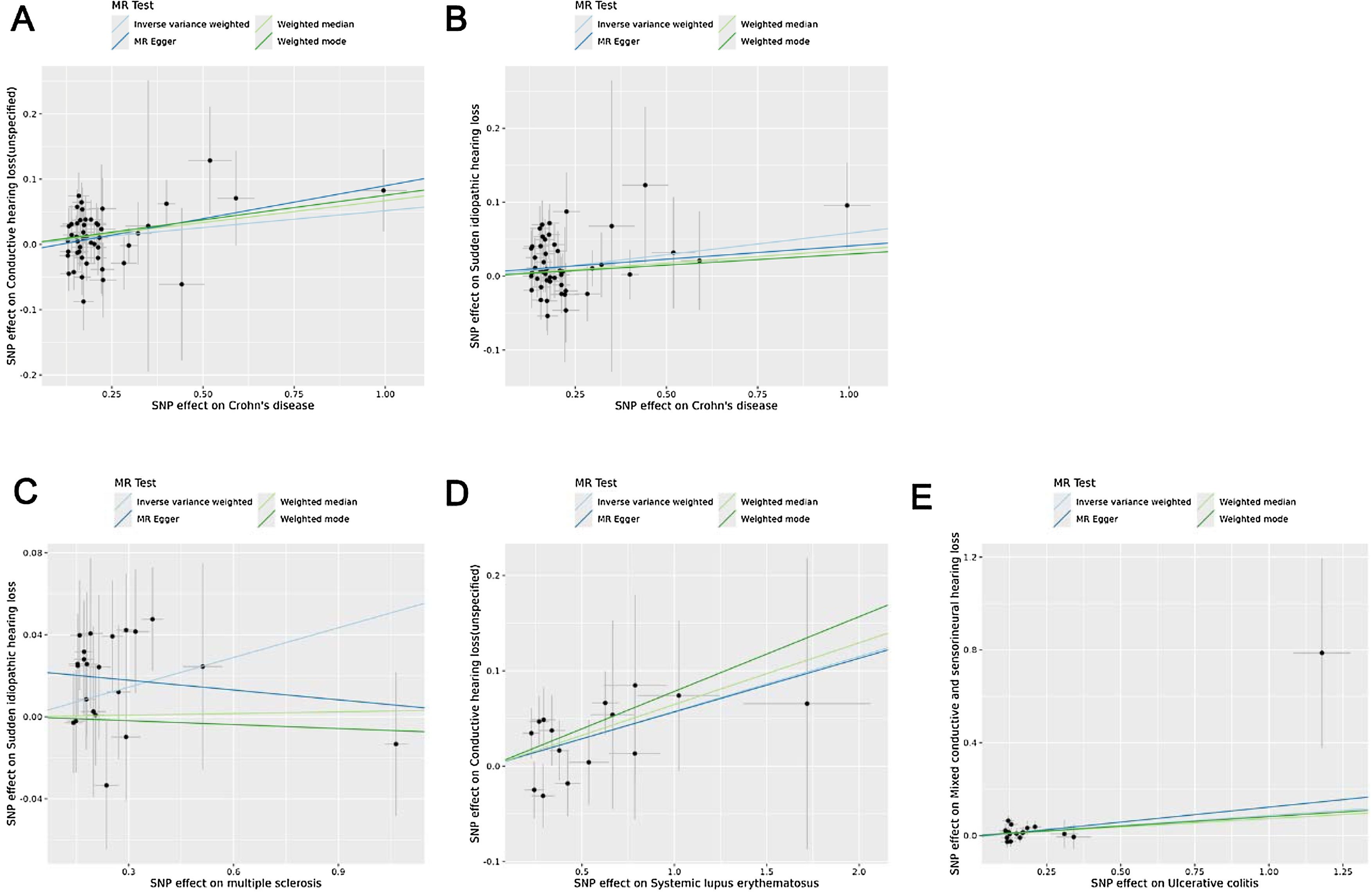
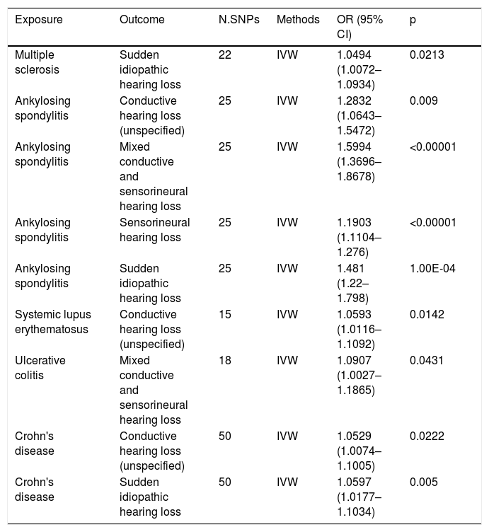
MR analysisMR analysis using the IVW method revealed nine significant AD–HL associations, including MS-SIHL (OR = 1.0494, 95% CI 1.0072–1.0934, p = 0.0213), AS-CHL (OR = 1.2832, 95% CI 1.0643–1.5472, p = 0.009), AS-MHL (OR = 1.5994, 95% CI 1.3696–1.8678, p < 0.00001), AS-SNHL (OR = 1.1903, 95% CI 1.1104–1.2760, p < 0.00001), AS-SIHL (OR = 1.481, 95% CI 1.22–1.798, p = 1.00E-04), SLE-CHL (OR = 1.0593, 95% CI 1.0116–1.1092, p = 0.0142), UC-MHL (OR = 1.0907, 95% CI 1.0027–1.1865, p = 0.0431), CD-CHL (OR = 1.0529, 95% CI 1.0074–1.1005, p = 0.0222), and CD-SIHL (OR = 1.0597, 95% CI 1.0177–1.1034, p = 0.005) (Table 1). The complete results of the MR analysis are shown in Table S5. Scatter plots (Figs. 2‒3 ) and forest plots (Fig. S1‒S2) illustrate these significant associations and SNP-specific effect estimates.
MR analysis of autoimmune diseases and hearing loss subtypes using the IVW method.
| Exposure | Outcome | N.SNPs | Methods | OR (95% CI) | p |
|---|---|---|---|---|---|
| Multiple sclerosis | Sudden idiopathic hearing loss | 22 | IVW | 1.0494 (1.0072–1.0934) | 0.0213 |
| Ankylosing spondylitis | Conductive hearing loss (unspecified) | 25 | IVW | 1.2832 (1.0643–1.5472) | 0.009 |
| Ankylosing spondylitis | Mixed conductive and sensorineural hearing loss | 25 | IVW | 1.5994 (1.3696–1.8678) | <0.00001 |
| Ankylosing spondylitis | Sensorineural hearing loss | 25 | IVW | 1.1903 (1.1104–1.276) | <0.00001 |
| Ankylosing spondylitis | Sudden idiopathic hearing loss | 25 | IVW | 1.481 (1.22–1.798) | 1.00E-04 |
| Systemic lupus erythematosus | Conductive hearing loss (unspecified) | 15 | IVW | 1.0593 (1.0116–1.1092) | 0.0142 |
| Ulcerative colitis | Mixed conductive and sensorineural hearing loss | 18 | IVW | 1.0907 (1.0027–1.1865) | 0.0431 |
| Crohn's disease | Conductive hearing loss (unspecified) | 50 | IVW | 1.0529 (1.0074–1.1005) | 0.0222 |
| Crohn's disease | Sudden idiopathic hearing loss | 50 | IVW | 1.0597 (1.0177–1.1034) | 0.005 |

Scatter plots show genetic associations between AS and HL subtypes identified in the primary MR analysis. (A) AS association with CHL (unspecified); (B) AS association with MHL; (C) AS association with SNHL; (D) AS association with SIHL. Regression lines for IVW, MR-Egger, weighted median, and weighted mode methods are included.

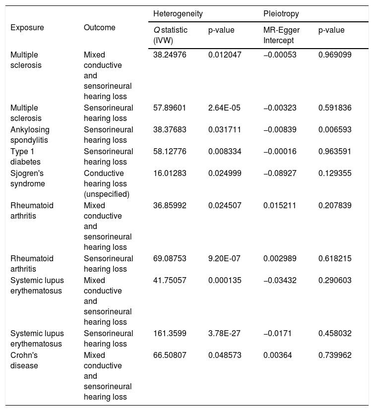
Significant heterogeneity was observed in ten AD-HL associations, including MS-MHL, MS-SNHL, AS-SNHL, T1D-SNHL, SS-CHL, RA-MHL, RA-SNHL, SLE-MHL, SLE-SNHL, and CD-MHL, with Q-statistic ranging from 16.01 to 161.36 (all p < 0.05). MR-Egger regression analysis identified significant horizontal pleiotropy in AS-SNHL association (MR-Egger intercept = −0.00839, p = 0.006593; Table 2). The complete results of the Heterogeneity and pleiotropy analysis analysis are shown in Table S6. No Heterogeneity and pleiotropy were observed in other significant IVW associations. Funnel plots illustrated horizontal pleiotropy (Fig. S3‒S4). Leave-one-out plots assessed the impact of individual IVs on overall MR estimates (Fig. S5‒S6).
Heterogeneity and horizontal pleiotropy in disease-hearing loss associations.
| Exposure | Outcome | Heterogeneity | Pleiotropy | ||
|---|---|---|---|---|---|
| Q statistic (IVW) | p-value | MR-Egger Intercept | p-value | ||
| Multiple sclerosis | Mixed conductive and sensorineural hearing loss | 38.24976 | 0.012047 | −0.00053 | 0.969099 |
| Multiple sclerosis | Sensorineural hearing loss | 57.89601 | 2.64E-05 | −0.00323 | 0.591836 |
| Ankylosing spondylitis | Sensorineural hearing loss | 38.37683 | 0.031711 | −0.00839 | 0.006593 |
| Type 1 diabetes | Sensorineural hearing loss | 58.12776 | 0.008334 | −0.00016 | 0.963591 |
| Sjogren's syndrome | Conductive hearing loss (unspecified) | 16.01283 | 0.024999 | −0.08927 | 0.129355 |
| Rheumatoid arthritis | Mixed conductive and sensorineural hearing loss | 36.85992 | 0.024507 | 0.015211 | 0.207839 |
| Rheumatoid arthritis | Sensorineural hearing loss | 69.08753 | 9.20E-07 | 0.002989 | 0.618215 |
| Systemic lupus erythematosus | Mixed conductive and sensorineural hearing loss | 41.75057 | 0.000135 | −0.03432 | 0.290603 |
| Systemic lupus erythematosus | Sensorineural hearing loss | 161.3599 | 3.78E-27 | −0.0171 | 0.458032 |
| Crohn's disease | Mixed conductive and sensorineural hearing loss | 66.50807 | 0.048573 | 0.00364 | 0.739962 |
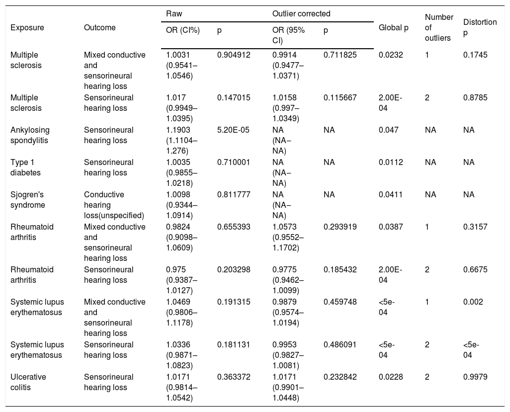
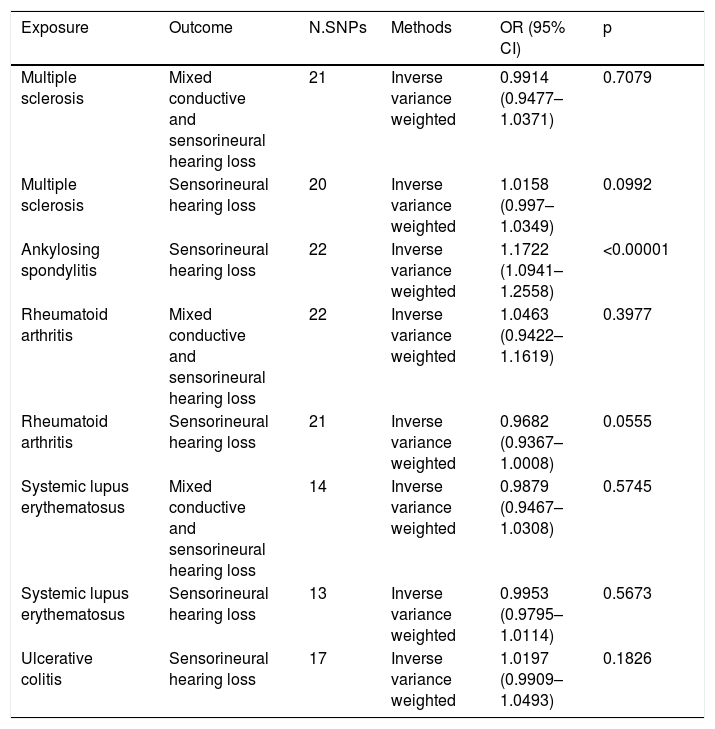
MR-PRESSO identified outliers in ten associations, including MS-MHL, MS-SNHL, AS-SNHL, T1D-SNHL, SS-CHL, RA-MHL, RA-SHL, SLE-MHL, SLE-SNHL, and UC-SNHL associations (Table 3). The complete results of the MR-PRESSO analysis analysis are shown in Table S7. Among these, only AS-SNHL exhibited a significant association in the initial IVW analysis. After removing outliers (except for T1D-SNHL and SS-CHL with unavailable outlier numbers), IVW results remained significant for AS-SNHL (OR = 1.1722, 95% CI 1.0941–1.2558, p < 0.00001), and no additional significant associations were found (Table 4). The MR analysis results after outlier removal are shown in Table S8.
Outlier detection and horizontal pleiotropy assessment using MR-PRESSO.
| Exposure | Outcome | Raw | Outlier corrected | Global p | Number of outliers | Distortion p | ||
|---|---|---|---|---|---|---|---|---|
| OR (CI%) | p | OR (95% CI) | p | |||||
| Multiple sclerosis | Mixed conductive and sensorineural hearing loss | 1.0031 (0.9541–1.0546) | 0.904912 | 0.9914 (0.9477–1.0371) | 0.711825 | 0.0232 | 1 | 0.1745 |
| Multiple sclerosis | Sensorineural hearing loss | 1.017 (0.9949–1.0395) | 0.147015 | 1.0158 (0.997–1.0349) | 0.115667 | 2.00E-04 | 2 | 0.8785 |
| Ankylosing spondylitis | Sensorineural hearing loss | 1.1903 (1.1104–1.276) | 5.20E-05 | NA (NA‒NA) | NA | 0.047 | NA | NA |
| Type 1 diabetes | Sensorineural hearing loss | 1.0035 (0.9855–1.0218) | 0.710001 | NA (NA‒NA) | NA | 0.0112 | NA | NA |
| Sjogren's syndrome | Conductive hearing loss(unspecified) | 1.0098 (0.9344–1.0914) | 0.811777 | NA (NA‒NA) | NA | 0.0411 | NA | NA |
| Rheumatoid arthritis | Mixed conductive and sensorineural hearing loss | 0.9824 (0.9098–1.0609) | 0.655393 | 1.0573 (0.9552–1.1702) | 0.293919 | 0.0387 | 1 | 0.3157 |
| Rheumatoid arthritis | Sensorineural hearing loss | 0.975 (0.9387–1.0127) | 0.203298 | 0.9775 (0.9462–1.0099) | 0.185432 | 2.00E-04 | 2 | 0.6675 |
| Systemic lupus erythematosus | Mixed conductive and sensorineural hearing loss | 1.0469 (0.9806–1.1178) | 0.191315 | 0.9879 (0.9574–1.0194) | 0.459748 | <5e-04 | 1 | 0.002 |
| Systemic lupus erythematosus | Sensorineural hearing loss | 1.0336 (0.9871–1.0823) | 0.181131 | 0.9953 (0.9827–1.0081) | 0.486091 | <5e-04 | 2 | <5e-04 |
| Ulcerative colitis | Sensorineural hearing loss | 1.0171 (0.9814–1.0542) | 0.363372 | 1.0171 (0.9901–1.0448) | 0.232842 | 0.0228 | 2 | 0.9979 |
MR analysis of disease-hearing loss associations post-outlier removal.
| Exposure | Outcome | N.SNPs | Methods | OR (95% CI) | p |
|---|---|---|---|---|---|
| Multiple sclerosis | Mixed conductive and sensorineural hearing loss | 21 | Inverse variance weighted | 0.9914 (0.9477–1.0371) | 0.7079 |
| Multiple sclerosis | Sensorineural hearing loss | 20 | Inverse variance weighted | 1.0158 (0.997–1.0349) | 0.0992 |
| Ankylosing spondylitis | Sensorineural hearing loss | 22 | Inverse variance weighted | 1.1722 (1.0941–1.2558) | <0.00001 |
| Rheumatoid arthritis | Mixed conductive and sensorineural hearing loss | 22 | Inverse variance weighted | 1.0463 (0.9422–1.1619) | 0.3977 |
| Rheumatoid arthritis | Sensorineural hearing loss | 21 | Inverse variance weighted | 0.9682 (0.9367–1.0008) | 0.0555 |
| Systemic lupus erythematosus | Mixed conductive and sensorineural hearing loss | 14 | Inverse variance weighted | 0.9879 (0.9467–1.0308) | 0.5745 |
| Systemic lupus erythematosus | Sensorineural hearing loss | 13 | Inverse variance weighted | 0.9953 (0.9795–1.0114) | 0.5673 |
| Ulcerative colitis | Sensorineural hearing loss | 17 | Inverse variance weighted | 1.0197 (0.9909–1.0493) | 0.1826 |
After outlier removal, significant heterogeneity persisted for MS-SNHL (Q = 35.36663, p = 0.012612), while both heterogeneity and horizontal pleiotropy were resolved for AS-SNHL (Table S9). In the MR-PRESSO analysis, the global p-value for MS-SNHL was less than 0.05, indicating potential pleiotropy. However, the number of outliers was unavailable, and the association was not influenced by horizontal pleiotropy. For all other analyses, neither horizontal pleiotropy nor significant heterogeneity was detected (Table S10).
DiscussionThis study provides evidence of causal relationships between ADs and HLs using MR analysis. Notable associations included AS-SNHL, AS-MHL, AS-CHL, AS-SIHL, SLE-CHL, UC-MHL, MS-SIHL, and CD-CHL and CD-SIHL. These results highlight the role of immune dysregulation in the pathogenesis of HL.
The association between AS and multiple HL subtypes highlights its potential role in auditory dysfunction. AS is a chronic inflammatory disease primarily affecting the axial skeleton, characterized by excessive immune activation, pro-inflammatory cytokine release, and systemic inflammation, which can contribute to vascular damage and multisystem involvement, including the auditory system.28 A systematic review and meta-analysis reported a 42.4% prevalence of HL in patients with AS, with significantly higher odds of HL and impaired hearing thresholds compared to individuals without AS.29 HL occurs when damage to the auditory system disrupts the function of inner ear cells and associated neural structures due to inflammation, oxidative stress, or vascular impairment.30 Pro-inflammatory cytokines elevated in AS, such as TNF-α and IL-6,31 disrupt cochlear microcirculation, leading to ischemic damage and impaired auditory function.32 Chronic inflammation in AS increases oxidative stress,33 which damages inner ear cells and contributes to HL. Vascular abnormalities frequently associated with AS, such as vasculitis and endothelial dysfunction,34 compromise blood flow to the cochlea, exacerbating auditory damage.35 These mechanisms provide a plausible explanation for the broad AS-HL associations observed in our study. Clinically, the robust AS-HL association warrants attention to auditory function in AS patients, particularly those with severe systemic inflammation or prolonged disease duration. Routine auditory monitoring may help identify early signs of HL and mitigate its impact on quality of life.
Following AS, Inflammatory Bowel Diseases (IBD), including CD and UC, were the second most frequently associated ADs with HL, demonstrating significant relationships with MHL, CHL, and SIHL. HL associated with CD may result from neuropathy of the cochlear nerve due to vitamin B1 deficiency, a common complication in CD caused by malnutrition, vomiting, or chronic inflammation.36 Additionally, SNHL in UC may be driven by autoimmune inner ear disease, immune-mediated damage to the cochlea, systemic inflammation, or ototoxic effects of medications commonly used in IBD treatment.37 Furthermore, mutations in the STXBP3 gene, implicated in very early onset IBD, disrupt epithelial cell polarization and immune regulation, which may contribute to bilateral SNHL in IBD, highlighting a genetic component to this association.38 Subclinical cochlear involvement, often beginning at high frequencies, has been documented in pediatric IBD patients and is proposed to result from vasculitis and immune complex deposition in cochlear vessels as additional mechanisms for HL.39 These findings underscore the importance of implementing early auditory screening and monitoring strategies in IBD patients with pediatric onset or severe systemic inflammation to prevent or mitigate HL progression effectively.
Significant associations were also found for SLE-CHL and MS-SIHL. SLE causes HL predominantly through immune complex deposition in the stria vascularis and endolymphatic sacs, leading to ischemia and cytotoxic damage to cochlear hair cells, with secondary SS further exacerbating the risk.40 Similarly, MS-SIHL may result from neuroinflammatory and neurodegenerative processes characteristic of MS, including oxidative stress, mitochondrial dysfunction, and demyelination within auditory pathways. The accumulation of toxic tau proteins in MS further disrupts neuronal function, potentially impairing auditory signal transmission and contributing to SNHL.41 Both SLE and MS involve inflammatory mechanisms that impair cochlear function, highlighting the need for targeted approaches to manage HL in these conditions.
This study provides novel evidence of robust AD-HL associations using MR methods. However, the relatively smaller sample sizes for certain conditions, such as SLE and less common HL subtypes may reduce statistical power. Additionally, the possibility of residual pleiotropy and confounding remains despite sensitivity analyses. The lack of longitudinal data further limits the ability to evaluate the temporal dynamics and potential reversibility of HL. Addressing these issues in future studies through larger datasets, refined statistical approaches, and longitudinal designs will enhance the robustness and clinical applicability of the findings.
ConclusionThis study identified significant causal associations between ADs and specific subtypes of HL. These findings underscore the role of immune dysregulation in the pathogenesis of auditory dysfunction and highlight the need for early audiological assessment and targeted therapeutic strategies in AD management to mitigate HL and improve patient outcomes.
ORCID IDYutong Huang: 0000-0002-0906-687X
Shengli Gao: 0000-0002-3523-2333
Renzhong Luo: 0000-0003-1521-4602
Authors’ contributionsYutong Huang carried out the studies, participated in collecting data, performed the statistical analysis and drafted the manuscript. Renzhong Luo and Shengli Gao participated in its design. All authors read and approved the final manuscript.
FundingThis study was supported by supported by scientific research foundation for Ph.D of Guangzhou Women and Children's Medical Center, Guangzhou Medical University (2021BS027, Applications of Electrically Evoked Cortical Auditory Evoked Potentials in Cochlear Implant), basci and applied basic research program of Guangzhou Municipal Science and Technology Bureau (2023A04J1240, Applications of Electrically Evoked Cortical Auditory Evoked Potentials in Postoperative Evaluation for Pediatric Cochlear), and Science and Technology Project of Guangzhou (2023A03J0909, etiology and genetic analysis of severe sensorineural hearing loss in children in the Pearl River Delta).
Conflicts of interestThe authors declare that the research was conducted in the absence of any commercial or financial relationships that could be construed as a potential conflict of interest.
Data availability statementAll data generated or analysed during this study are included in this published article and its Supplementary Information File.
Not applicable.
The following is Supplementary data to this article:




