





To compare the diagnostic performance of 18F-fluorodeoxyglucose (18F-FDG) and 18F-PSMA-1007 PET/CT in patients with locoregionally advanced head and neck squamous cell carcinoma (HNSCC).
MethodsFourteen patients with HNSCC meeting locoregionally advanced criteria at PET/CT (stage III–IVB at presentation and/or locoregionally advanced recurrence/progression) underwent 18F-FDG and 18F-PSMA-1007 PET/CT from September 2023 to December 2024; imaging was performed at diagnosis or locoregional recurrence, and one had a stage II primary imaged during locoregionally advanced recurrence. Detection rates of primary and metastatic lesions and standardized uptake values (SUVmax) were assessed and compared between tracers.
Results18F-FDG PET/CT identified all primary lesions and metastatic lesions, whereas 18F-PSMA-1007 showed uptake in 10/13 evaluable primary tumors (76.9%) and in 6/13 patients with metastatic disease (46.2%). The median SUVmax values of primary tumors at 1 h and 2 h post-injection were significantly higher with 18F-FDG PET/CT compared to 18F-PSMA-1007 PET/CT (21.0 [IQR = 19.1] vs. 4.3 [IQR = 2.7] and 24.2 [IQR = 17.4] vs. 5.8 [IQR = 2.3], respectively; p < 0.001). In larger tumors, both tracers exhibited distinct intralesional uptake patterns, suggesting different biological targets.
Conclusion18F-FDG PET/CT demonstrated superior lesion detection compared to 18F-PSMA-1007 PET/CT in advanced HNSCC. However, the complementary distribution of both tracers indicates that glycolytic activity and neovascular PSMA expression may coexist in tumors. These findings highlight the potential role of 18F-PSMA-1007 PET/CT as an adjunct to 18F-FDG PET/CT for the biological characterization of HNSCC, particularly in identifying angiogenic regions that may not be captured by conventional metabolic imaging.
Level of evidence4.
Head and neck squamous cell carcinoma (HNSCC) is recognized as the eighth most prevalent cancer worldwide, accounting for approximately 771,307 new cases and 384,631 deaths annually.1 The incidence of HNSCC is higher among males, tobacco users, and individuals with a history of alcohol abuse,2,3 as well as among individuals infected with human papillomavirus (HPV).3–5
In clinical practice, therapeutic management of HNSCC is determined by the primary tumor site and staging.6–8 Staging and follow-up of HNSCC are traditionally performed through computed tomography (CT).918F-fluorodeoxyglucose (18F-FDG), a marker of glycolytic activity, has been used in positron emission tomography/computed tomography (18F-FDG PET/CT), which enables comprehensive evaluation of locoregional disease extent and distant metastases.8,10 Surgical resection (SR) and radiotherapy (RT) represent the main approaches for patients with localized disease,11 while patients with metastatic disease require platinum-based chemotherapy,12–14 and multimodal treatment is indicated for patients with locally advanced tumors.7,15
Despite the above-mentioned procedures, the overall survival (OS) rate for patients with HNSCC remains around 40%–50% at five-years,16,15 highlighting the need for improved strategies for tumor characterization, staging, treatment, and treatment monitoring.
Prostate-specific membrane antigen (PSMA), a marker of neoangiogenesis in tissues, was initially recognized for its overexpression in prostate cancer cells.17,18 Gallium-68 PSMA PET/CT scans have been used for detecting metastases of prostate cancer,19 and lutetium-177 PSMA has been indicated for treatment of castration-resistant metastatic prostate cancer.20 Previous studies have demonstrated PSMA expression in non-prostatic malignancies. Incidental detection of lung, lymphoma, thyroid, and base-of-tongue primary malignancies was identified by 68Ga-PSMA PET/CT in patients with prostate cancer.21 Renal cell carcinoma, breast cancer, hepatocellular carcinoma, and glioblastoma were also identified by PSMA PET/CT.22–28 PSMA uptake was observed in adenoid cystic carcinoma, salivary duct carcinoma,29 and concomitant prostate cancer and SCC of the oropharynx30 and larynx31 cases, but the clinical role of PSMA PET/CT in HNSCC remains undefined. In this context, the current study aimed to investigate the role of PSMA PET/CT in the characterization of HNSCC, considering 18F-FDG PET/CT as the reference method among imaging modalities.
We hypothesized that, in HNSCC, 18F-PSMA-1007 PET/CT may provide biologically distinct information from 18F-FDG PET/CT by preferentially reflecting tumor-associated neoangiogenesis, whereas FDG predominantly reflects glycolytic metabolism within the tumor microenvironment. Therefore, both tracers may have complementary clinical value for lesion characterization and for depicting intratumoral and interlesional heterogeneity.
MethodsPatientsThis cross-sectional observational study was conducted at the Clinical Oncology Service of the General Hospital of the University of Campinas from September 4, 2023, to December 12, 2024.
Inclusion criteria were age >18-years, histopathological diagnosis of HNSCC involving the oral cavity, pharynx, or larynx, Eastern Cooperative Oncology Group (ECOG) performance status ≤2 and locoregionally advanced disease at the time of PET/CT, defined as AJCC 8th edition stage III–IVB (M0) at presentation and/or locoregionally advanced locoregional recurrence/progression requiring multimodality management. One patient with a prior stage II primary was included because imaging was performed in the setting of locoregionally advanced recurrence. All participants provided written informed consent. Exclusion criteria included nasopharyngeal tumors, pregnancy, prior history of another malignant neoplasm, severe comorbidities, insufficient medical records, and refusal to participate in the study.
For the purposes of this study, the term locoregionally advanced HNSCC refers to non-metastatic disease (M0) evaluated either at initial diagnosis or at locoregional recurrence. When present, distant lesions were recorded as metastatic sites for detection-rate analysis; however, metastatic disease was not a separate inclusion stratum.
The confirmation of HNSCC at diagnosis, relapse and metastasis, and the identification of tumor grading were based on histological exam, in accordance with World Health Organization (WHO) criteria.32 Tumor staging was defined using the the American Joint Committee on Cancer (AJCC) criteria.6
Imaging examinationsEach patient underwent two imaging examinations: an 18F-FDG PET/CT and an 18F-PSMA-1007 PET/CT. Both scans were performed within a 24h interval to ensure temporal proximity, allowing for a detailed comparison between glycolytic metabolism and neoangiogenesis status in tumor lesions.
Prior to the injection of 18F-FDG, patients fasted for at least six hours and underwent stimulated hydration. Additionally, serum glucose levels were confirmed to be below 200 mg/dL.33 In contrast, no specific preparation was required prior to the administration of 18F-PSMA-1007. FDG and PSMA PET/CT images were acquired 60 and 120 minutes after the intravenous administration of 0.10 mCi/kg 18F-FDG and 0.11 mCi/kg of 18F-PSMA-1007, covering the region from the vertex to the thighs, using a dedicated PET/CT scanner (Biograph mCT40, Siemens Healthcare, USA). Furthermore, all patients underwent delayed imaging two hours after injection, specifically targeting the head and neck region. CT parameters included 5 mm axial reconstruction with automatic dose modulation, and PET acquisitions were performed in 3D mode with a duration of 90 seconds per bed position. Images were reconstructed and displayed in axial, coronal, and sagittal planes.
The PET/CT images were independently analyzed by two board-certified nuclear medicine physicians. PET/CT findings were interpreted by visual analysis integrated with CT. A lesion was classified as positive when it showed focal uptake clearly higher than the uptake in surrounding normal tissues (adjacent physiological uptake), not explained by physiological distribution, with or without a corresponding suspicious CT abnormality. A lesion was classified as negative when uptake was absent or compatible with physiological activity and there was no suspicious CT correlate. Findings were considered indeterminate when uptake was mild/equivocal and/or when CT findings were nonspecific, such as small lymph nodes without definitive malignant features or inflammatory-appearing changes. Discrepancies between readers were resolved by consensus.
The reference standard for lesion status was established by a combined analysis of pathological and clinical data. For the primary tumor, positivity was defined by histopathological confirmation (biopsy or surgical specimen), in accordance with WHO criterio.32 For metastatic lesions, histopathology was used whenever available. When biopsy was not feasible, lesions were classified as true metastases based on clinical-radiological follow-up, defined as typical malignant behavior on serial imaging (progressive increase in size/number, appearance of new lesions in expected metastatic sites) and/or a concordant response pattern to oncologic treatment. Lesions without histological confirmation and without unequivocal progression/response on follow-up were considered unconfirmed.
Quantitative interpretation (MFS VB20, syngo.via MM Oncology, Siemens Medical Solutions, USA) was conducted on all PET/CT images to determine total body tumor burden. FDG and PSMA uptake in tumor lesions was quantified using the maximum standardized uptake value (SUVmax). Coregistration of FDG PET/CT and PSMA PET/CT images ensured accurate overlap of functional and morphological data. Combined analysis of FDG PET/CT and PSMA PET/CT findings with clinical and pathological data was conducted by the study’s principal investigator and the nuclear medicine physicians, ensuring a comprehensive and integrated assessment. The SUVmax of the primary tumor and the metastatic site lesions were assessed for both radiopharmaceuticals (FDG and PSMA). The total numbers of radiotracer-avid lesions were also analyzed individually for each radiotracer and for lesions demonstrating uptake with both radiotracers simultaneously.
Statistical analysisData were collected through a semi-structured questionnaire, tabulated and organized using Microsoft Office Excel version 2016, and analyzed using the JASP software (version 0.18.3). Descriptive statistics were expressed as mean, median, standard deviation, interquartile range, and range, as appropriate. Given the small sample size and the non-parametric distribution of the dataset, comparisons between groups were performed using the Mann–Whitney U-test. A p-value <0.05 was considered statistically significant.
Ethical considerationsThe study was approved by the Institutional Research Ethics Committee under approval number 30476720.9.0000.5404, in accordance with international ethical guidelines for research involving human subjects. All patients included in the study provided written informed consent after receiving detailed explanations regarding the study’s objectives, procedures, potential risks, and benefits, ensuring their voluntary and informed participation.
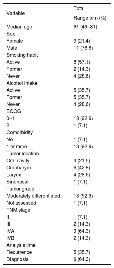
ResultsThe demographic and clinicopathological characteristics of the 14 patients included in the study are presented in Table 1. The median age was 61-years, with a predominance of male, smokers, and alcoholic patients. Most patients had good functional performance, one or more comorbidities, moderately differentiated tumors, and predominantly AJCC 8th edition stage III–IVB disease (M0) at presentation; one patient had a prior stage II primary but was imaged during locoregionally advanced recurrence. The tumors were located predominately in the oropharynx, larynx or oral cavity. Nine patients were analyzed at diagnosis and five patients at disease recurrence.
Sociodemographic and clinical pathological characteristics of patients with head and neck squamous cell carcinoma analyzed by positron emission tomography/computed tomography.
| Variable | Total |
|---|---|
| Range or n (%) | |
| Median age | 61 (49–81) |
| Sex | |
| Female | 3 (21.4) |
| Male | 11 (78.6) |
| Smoking habit | |
| Active | 8 (57.1) |
| Former | 2 (14.3) |
| Never | 4 (28.6) |
| Alcohol intake | |
| Active | 5 (35.7) |
| Former | 5 (35.7) |
| Never | 4 (28.6) |
| ECOG | |
| 0‒1 | 13 (92.9) |
| 2 | 1 (7.1) |
| Comorbidity | |
| No | 1 (7.1) |
| 1 or more | 13 (92.9) |
| Tumor location | |
| Oral cavity | 3 (21.5) |
| Oropharynx | 6 (42.8) |
| Larynx | 4 (28.6) |
| Sinonasal | 1 (7.1) |
| Tumor grade | |
| Moderately differentiated | 13 (92.9) |
| Not assessed | 1 (7.1) |
| TNM stage | |
| II | 1 (7.1) |
| III | 2 (14.3) |
| IVA | 9 (64.3) |
| IVB | 2 (14.3) |
| Analysis time | |
| Recurrence | 5 (35.7) |
| Diagnosis | 9 (64.3) |
ECOG, Eastern Cooperative Oncology Group; TNM, tumor-lymph node-metastasis classified by American Joint Committee on Cancer (AJCC) criteria.
One patient with a prior AJCC 8th edition stage II primary was included because imaging was performed during stage III recurrence (per inclusion criteria).
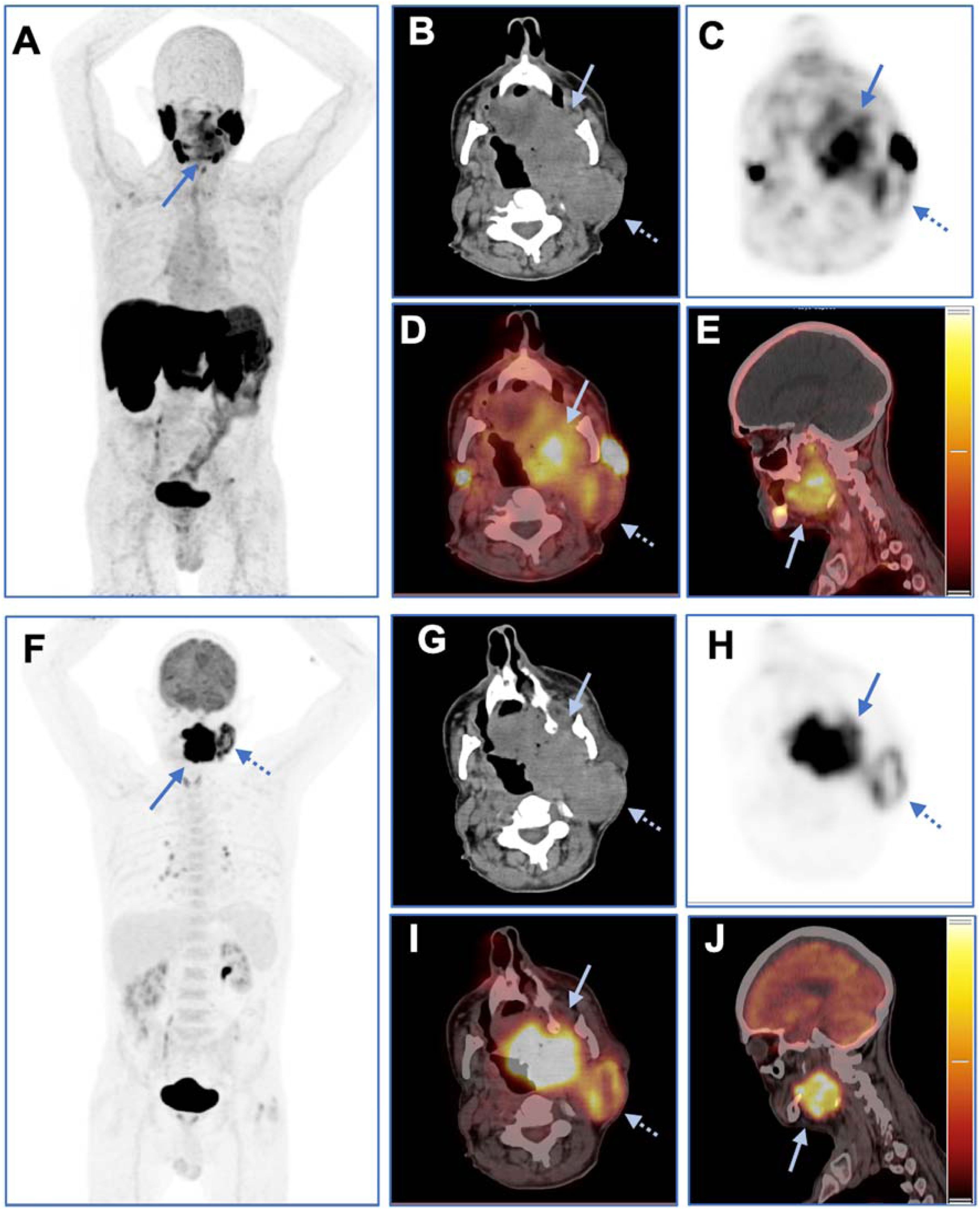
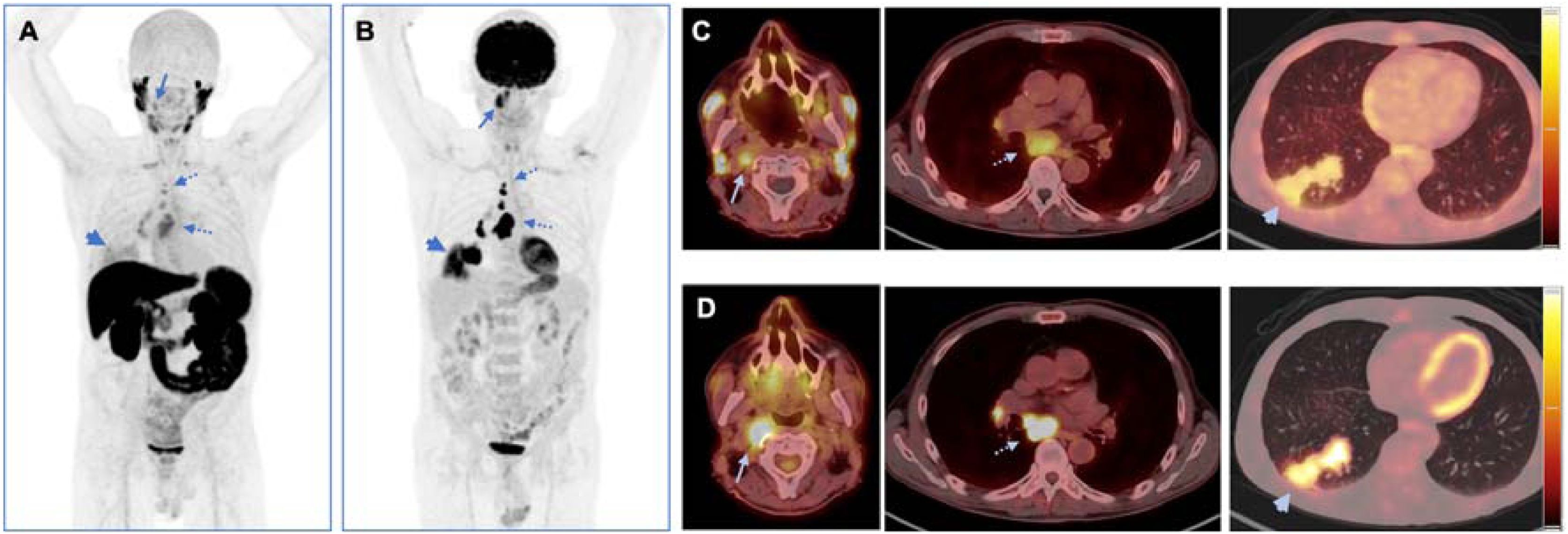
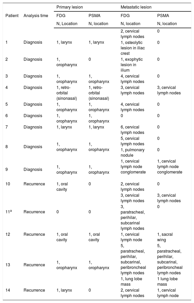
The numbers of primary and metastatic lesions detected by FDG PET/CT and PSMA PET/CT are presented in Table 2 and Supplementary Table S1. Excluding patient 11, who had undergone total laryngectomy and therefore lacked a primary anatomical site for evaluation, FDG PET/CT detected primary lesions in all 13 evaluable patients (100.0%), whereas PSMA PET/CT demonstrated uptake in 10 of the 13 patients (76.9%). All primary lesions detected by PSMA PET/CT were also detected by FDG PET/CT (Fig. 1). Excluding patient 6, who presented only locally advanced disease, FDG PET/CT identified metastases in cervical lymph nodes, pulmonary lymph nodes, pulmonary parenchyma, and bone in 13 patients (100.0%), whereas 6 out of 13 patients (46.2%) presented metastases in cervical lymph nodes, pulmonary lymph nodes, and pulmonary parenchyma detected by PSMA PET/CT (Fig. 2). All metastatic lesions detected by PSMA PET/CT were also detected by FDG PET/CT, except an unbiopsied sclerotic lesion in the sacral wing in patient 12, which was seen only by PSMA PET/CT.
Lesions detected by positron emission tomography/computed tomography in patients with head and neck squamous cell carcinoma.
| Patient | Analysis time | Primary lesion | Metastatic lesion | ||
|---|---|---|---|---|---|
| FDG | PSMA | FDG | PSMA | ||
| N, Location | N, location | N, location | N, location | ||
| 1 | Diagnosis | 1, larynx | 1, larynx | 2, cervical lymph nodes | 0 |
| 1, osteolytic lesion in iliac crest | 0 | ||||
| 2 | Diagnosis | 1, oropharynx | 0 | 1, exophytic lesion in ilium | 0 |
| 3 | Diagnosis | 1, oropharynx | 1, oropharynx | 4, cervical lymph nodes | 0 |
| 4 | Diagnosis | 1, retro-orbital (sinonasal) | 1, retro-orbital (sinonasal) | 3, cervical lymph nodes | 3, cervical lymph nodes |
| 5 | Diagnosis | 1, oropharynx | 1, oropharynx | 4, cervical lymph nodes | 0 |
| 6 | Diagnosis | 1, oropharynx | 1, oropharynx | 0 | 0 |
| 7 | Diagnosis | 1, larynx | 1, larynx | 6, cervical lymph nodes | 0 |
| 8 | Diagnosis | 1, oropharynx | 1, oropharynx | 5, cervical lymph nodes | 0 |
| 1, pulmonary nodule | 0 | ||||
| 9 | Diagnosis | 1, oropharynx | 1, oropharynx | 1, cervical lymph node conglomerate | 1, cervical lymph node conglomerate |
| 10 | Recurrence | 1, oral cavity | 0 | 2, cervical lymph nodes | 0 |
| 11a | Recurrence | 0 | 0 | 3, cervical lymph nodes | 3, cervical lymph nodes |
| 3, paratracheal, perihilar, subcarinal lymph nodes | 0 | ||||
| 12 | Recurrence | 1, oral cavity | 1, oral cavity | 1, cervical lymph node | 1, sacral wing |
| 13 | Recurrence | 1, oropharynx | 1, oropharynx | 5, paratracheal, perihilar, subcarinal, peribroncheal lymph nodes | 5, paratracheal, perihilar, subcarinal, peribroncheal lymph nodes |
| 1, lung lobe mass | 1 lung lobe mass | ||||
| 14 | Recurrence | 1, larynx | 0 | 2, cervical lymph nodes | 1, cervical lymph node |
FDG, 18-fluorodeoxyglucose; PSMA, prostate-specific membrane antigen 1007.

PET/CT images with 18F-PSMA-1007 (top row, A–E) and 18F-FDG (bottom row, F–J) in a patient with squamous cell carcinoma involving the right oral cavity and oropharynx, associated with left-sided cervical lymphadenopathy (Patient 9). Focal radiotracer uptake is seen in the primary lesion site (solid arrows) and in a left cervical nodal conglomerate with central necrosis, centered in the parotid chain (dashed arrows). Axial CT (B and G), PET (C and H), fused PET/CT (D and I), and sagittal reconstructions (E and J) confirm the anatomical and functional findings. 18F-FDG PET/CT uptake was markedly higher in both the primary tumor (SUVmax1 h: 59.8 vs. 6.2; SUVmax2 h: 63.7 vs. 6.9) and the metastatic lymph node (SUVmax1 h: 15.8 vs. 3.9; SUVmax2 h: 22.0 vs. 4.2). Small mediastinal lymph nodes with FDG uptake (F), but no PSMA uptake (A), were observed, likely reflecting inflammatory changes rather than metastasis (a potential false-positive FDG finding).

Maximum Intensity Projection (MIP) of 18F-PSMA-1007 (A) and 18F-FDG (B) PET/CT images of a patient with right-sided oropharyngeal squamous cell carcinoma and mediastinal and pulmonary metastases (Patient 13). Axial slices of fused 18F-PSMA PET/CT images of the head and chest (soft tissue and pulmonary windows) are shown in (C), and the corresponding slices of 18F-FDG PET/CT are shown in (D). Lesion uptake of both tracers is seen in the primary site, located in the right pharyngeal tonsil (solid arrows), with more intense and extensive for 18F-FDG (SUVmax1 h: 10.7, SUVmax2 h: 12.9) than for 18F-PSMA-1007 (SUVmax90 min: 3.0, SUVmax2 h: 4.4). Additionally, multiple metastatic mediastinal lymph nodes, including upper and lower paratracheal, subcarinal, and right hilar chains are seen with both tracers (dashed arrows), with markedly higher FDG uptake (SUVmax2 h up to 34.4) than PSMA uptake (SUVmax2 h up to 6.9). A spiculated pulmonary mass is also identified in the posterior basal segment of the right lower lobe, with intense FDG uptake (SUVmax2 h: 25.9) and moderate PSMA uptake (SUVmax2 h: 15.7), consistent with pulmonary metastasis (arrowheads).
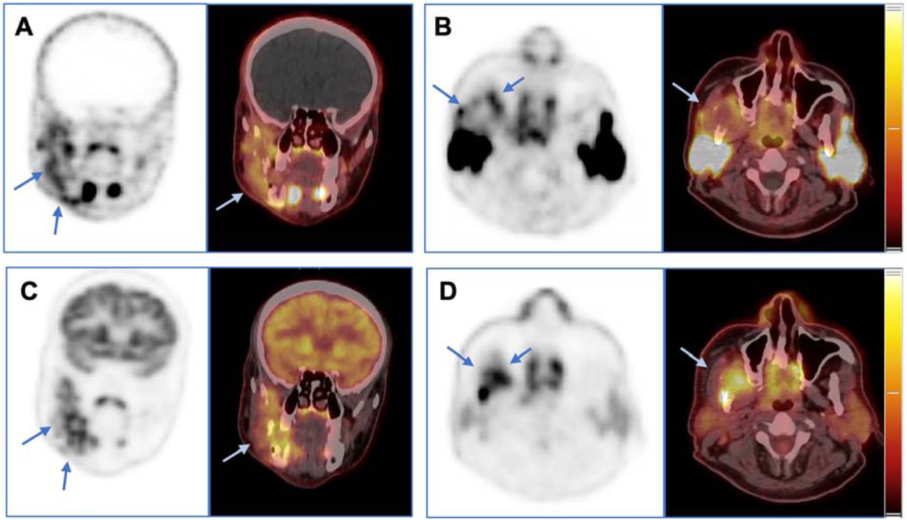
Ten out of 13 patients with primary tumors (76.9%), 6 out of 13 patients with metastatic disease (46.2%) were identified by both FDG PET/CT and PSMA PET/CT (Fig. 1–2). In patient 1 and patient 3, both radiotracers demonstrated concordant uptake in the same indeterminate lesions; in patient 6, FDG showed uptake in two lesions, while PSMA identified only one of them. Interestingly, patients with largest tumors presented intralesional patterns with marked differences (Fig. 3).

Coronal (A) and axial (B) slices showing PET (right) and fused PET/CT images (left) obtained with 18F-PSMA-1007 from a patient with squamous cell carcinoma of the right retromolar trigone, with bone invasion (Patient 6). The corresponding 18F-FDG images are shown in (C) and (D). Lesion uptake of both tracers is clearly seen in the ill-defined primary lesion (arrows). However, their intralesional patterns differ markedly: 18F-PSMA-1007 displays peripheral uptake (A and B), likely reflecting PSMA expression in the tumor's neovascular periphery, while 18F-FDG shows predominant central uptake (C and D), consistent with increased glycolytic activity in a hypoxic or partially necrotic tumor core.
In the analysis of FDG-positive/PSMA-negative findings, PSMA PET/CT showed no visually relevant uptake at the primary site in three cases, whereas FDG PET/CT identified the primary lesion in these patients. These PSMA-negative primaries involved the oropharynx (patient 2), oral cavity (patient 10), and larynx (patient 14). At the metastatic level, several FDG-avid deposits, predominantly cervical nodal lesions across levels II–V/VI, showed no corresponding PSMA uptake, underscoring lesion-dependent heterogeneity of PSMA targeting in this cohort. This description is based on lesion-level mapping (Supplementary Table S1) and was not subjected to formal statistical testing.
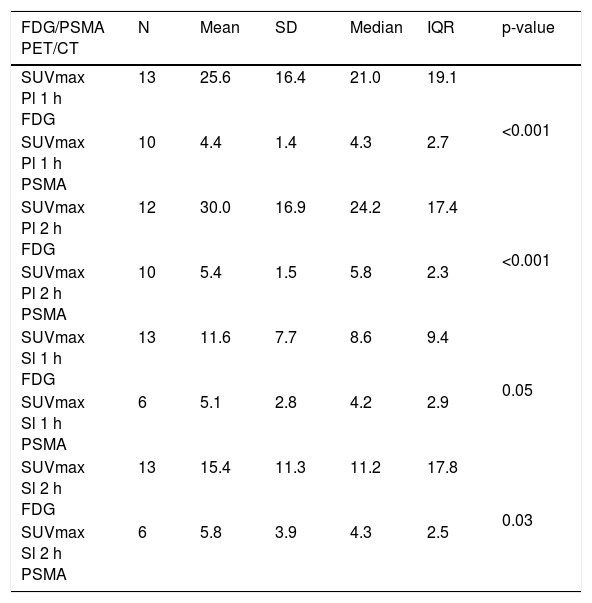
The maximum FDG and PSMA uptake values at 1h and 2h post-injection are presented in Table 3. The mean SUVmax values in primary lesions detected by FDG PET/CT at 1 and 2h were higher than those detected by PSMA PET/CT. For metastatic lesions, the mean SUVmax values detected by FDG PET/CT at 1 and 2h tended to be higher than those detected by PSMA PET/CT.
Positron emission tomography/computed tomography uptake in head and neck squamous cell carcinoma.
| FDG/PSMA PET/CT | N | Mean | SD | Median | IQR | p-value |
|---|---|---|---|---|---|---|
| SUVmax Pl 1 h FDG | 13 | 25.6 | 16.4 | 21.0 | 19.1 | <0.001 |
| SUVmax Pl 1 h PSMA | 10 | 4.4 | 1.4 | 4.3 | 2.7 | |
| SUVmax Pl 2 h FDG | 12 | 30.0 | 16.9 | 24.2 | 17.4 | <0.001 |
| SUVmax Pl 2 h PSMA | 10 | 5.4 | 1.5 | 5.8 | 2.3 | |
| SUVmax Sl 1 h FDG | 13 | 11.6 | 7.7 | 8.6 | 9.4 | 0.05 |
| SUVmax Sl 1 h PSMA | 6 | 5.1 | 2.8 | 4.2 | 2.9 | |
| SUVmax Sl 2 h FDG | 13 | 15.4 | 11.3 | 11.2 | 17.8 | 0.03 |
| SUVmax Sl 2 h PSMA | 6 | 5.8 | 3.9 | 4.3 | 2.5 |
FDG, fluorodeoxyglucose; PSMA, prostate-specific membrane antigen, PET/CT, positron emission tomography/computed tomography; Pl, primary lesion; SL, secondary lesion; N, number of patients analyzed; SUVmax, maximum standardized uptake value; SD, standard deviation; IQR, interquartile range.
The current study provides a head-to-head comparison of 18F-FDG and 18F-PSMA-1007 PET/CT in locoregionally advanced HNSCC, demonstrating that while FDG remains superior for tumor detection, PSMA offers complementary insights into tumor biology, particularly in characterizing angiogenesis and intratumoral heterogeneity.
It was observed that HNSCC lesions were more consistently identified by 18F-FDG PET/CT than by 18F-PSMA-1007 PET/CT, particularly for metastatic lesions, and 18F-FDG PET/CT outperformed 18F-PSMA-1007 PET/CT in sensitivity (100% vs. 76.9% for primary lesions; 100% vs. 46.2% for metastases).
Although PSMA uptake is biologically linked to tumor-associated neovasculature, the PSMA PET/CT detection rate observed in our cohort was lower than might be expected from the reported frequency of vascular PSMA expression in HNSCC histopathological series. In our study, 18F-PSMA-1007 demonstrated visually relevant uptake in the primary tumor in 10/13 evaluable patients (76.9%) and detected metastatic disease in 6/13 patients (46.2%), whereas FDG detected all evaluable primary and metastatic lesions. Tissue-based data have shown frequent, yet heterogeneous, endothelial PSMA expression in HNSCC, with a graded distribution of microvessel involvement and higher vascular PSMA expression in metastatic lymph nodes than in reactive nodes.34,35 Therefore, histologic vascular PSMA positivity does not necessarily translate into lesion detectability on PET, consistent with lesion-dependent heterogeneity of endothelial expression.
In fact, FDG PET/CT has assumed an important role in the initial assessment36 and early detection of recurrences in HNSCC.37,38 More recently, the diagnostic potential of PET/CT using radiotracers targeting PSMA, already established in prostate cancer, has been investigated in other solid tumors.22–29
In HNSCC, synchronous SCC (squamous cell carcinoma) of the oropharynx was identified in a retrospective study involving 764 patients with prostate cancer,21 an invasive SCC of the oropharynx was detected during prostate cancer staging,30 and, more recently, the use of a dual-tracer PET/CT approach with 18F-FDG and 18F-PSMA-1007 demonstrated FDG and PSMA uptake in both prostate and laryngeal carcinoma.31 Considering that PSMA uptake has been previously reported only in incidental cases of HNSCC, this is the first study that analyzed PET/CT using radiotracers targeting PSMA in a series of HNSCC patients. In addition, as 18F-FDG PET/CT enables the detection of glycolytic activity across the tumor microenvironment,8,10 and PSMA uptake reflects tumor neo vasculature,34,35 the results of the current study present, for the first time, preliminary evidence that metabolic reprogramming and neoangiogenesis are hallmarks of HNSCC.
It is worth commenting that one false positive finding was attributed to PSMA PET/CT. In patient 12, an increased PSMA uptake was observed in a unbiopsied sclerotic bone lesion located in the right sacral wing, without corresponding metabolic activity on FDG PET/CT or anatomical findings on CT. A high prevalence of nonspecific bone uptake on PSMA-1007 PET in patients with prostate cancer has been reported,39 and this finding was validated with follow-up PSMA PET/CT.40 Such nonspecific uptake may be attributed to PSMA expression in the neovasculature associated with inflammatory conditions and changes in vascular permeability.41
In the current study, it was observed that the mean SUVmax values in primary lesions detected by FDG PET/CT at 1 and 2h were higher than those detected by PSMA PET/CT, with an increase in both tracers’ uptake over time. In a review of PSMA-targeted radiopharmaceuticals in non-prostatic tumors, it was reported that SUVmax values observed on PSMA PET/CT were generally lower than those seen in prostate cancer, and that tracer distribution tended to be more heterogeneous.42 Although HNSCC was not included in that analysis, it was emphasized that in most of the evaluated tumors, PSMA expression was confined to the tumor neovasculature.43,44 In another report, when synchronous prostate and laryngeal carcinomas were evaluated by both tracers, the laryngeal carcinoma exhibited intense FDG uptake with only mild PSMA avidity, while the prostate carcinoma demonstrated the opposite pattern.31 The results of the current study support the concept that 18F-FDG PET/CT demonstrates more intense uptake and more diffuse distribution in HNSCC when compared to PSMA PET/CT.
Interestingly, it was also observed in the current study that patients with large lesions presented markedly different intralesional patterns, as in patient 6. FDG showed central uptake in the primary lesion, aligning with its affinity for hypoxic, glycolytically active tumor cores.45 PSMA exhibited peripheral uptake, likely due to PSMA expression in tumor-associated neovasculature.34 The central necrosis or hypoxia may limit PSMA expression to viable peripheral regions. This spatial dichotomy underscores the complementary roles of FDG (metabolic activity) and PSMA (angiogenesis) in HNSCC characterization. Similar findings have been reported in prostate cancer43 and glioblastoma,26 where PSMA uptake correlates with tumor microvessel density rather than epithelial proliferation. The distinct uptake patterns observed may have therapeutic implications: FDG’s central dominance supports its role in staging and surveillance, particularly for identifying aggressive, hypoxic tumor subclones resistant to radiotherapy.46 PSMA’s peripheral localization suggests potential utility for guiding anti-angiogenic therapies (e.g., VEGF inhibitors) in PSMA-avid subgroups and for selecting patients for PSMA-targeted radioligand therapy (e.g., 177Lu-PSMA), as proposed for other solid tumors.47
The current study has some limitations. The small sample size, both at initial diagnosis and at recurrence, and the single-center design may limit the statistical power and the generalizability of the findings. Another point is that the proliferative index Ki-67 as well as molecular characterization were not available for these patients, precluding correlation with the metabolic and/or angiogenic activity observed on imaging studies.
ConclusionIn conclusion, our data present preliminary evidence that 18F-FDG PET/CT is indispensable for HNSCC staging while 18F-PSMA-1007 provides complementary angiogenic profiling, contributing to tumor characterization. Further larger studies with patients at diagnosis and relapse from various services and with well tumor characterization have to be conducted to define consistent conclusions about the roles of PET/CT with 18F-FDG and 18F-PSMA-1007 in staging of HNSCC and in the selection of candidates for targeted therapies.
ORCID IDHádila da Silva Veras Sousa: 0009-0000-8400-9456
Victor Cabral Costa Ribeiro Heringer: 0000-0002-1649-5656
Matheus Yung Perin: 0000-0001-6981-9063
Nájua Abou Arabi Silveira: 0009-0003-5772-4288
Simone Kuba: 0009-0009-2414-7705
Ligia Traldi Macedo: 0000-0003-0575-586X
Gustavo Jacob Lourenço: 0000-0002-5944-0305
Carlos Takahiro Chone: 0000-0002-4217-4629
Carmino Antônio de Souza: 0000-0001-8656-8374
Celso Dario Ramos: 0000-0002-1312-1026
Carmen Silvia Passos Lima: 0000-0002-1314-2345
Justification for the number of authorsThe number of authors exceeds the journal’s limit of seven; however, we request the inclusion of all eleven authors due to their substantial and distinct contributions to the study. This work was developed by a multidisciplinary team that included oncologists, radiotherapists, and surgeons, whose expertise was essential for patient selection, data acquisition, and interpretation.
HSVS contributed to patient selection, data analysis, manuscript drafting, and critical revision. MYP and LTM contributed to patient selection. VCCRH, and SC were responsible for performing imaging examinations and conducting image analysis. GJL performed the statistical analyses. CTC, CAS, CDR and CSPL contributed to research planning, study design, manuscript drafting, and critical revision. All authors have read and approved the final version of the manuscript and agree to be accountable for all aspects of the work.
FundingWe are grateful for financial support from Fundação de Amparo a Pesquisa do Estado de São Paulo, Brazil, [Grant FAPESP #2021/10265-8], Cancer Theranostics Innovation Center/Centro de Pesquisa, Inovação e Difusão, CancerThera/CEPID; and National Council for Scientific and Technological Development, CNPq, Research Productivity, [Grant #307944/2022-0(Lourenço GJ)], [Grant #312212/2025-9(Ramos CD)], and [Grant #302922/2025-3 (Lima CSP)].
Data availability statementThe authors declare that all data are available in repository.
The authors declare no conflict of interest.
The following are Supplementary data to this article:




