




This study presents a case of asymptomatic Intrathyroidal Parathyroid Carcinoma (IPC) and a scoping review of the literature from the past 25-years, aiming to characterize the clinical and laboratory profiles and diagnostic difficulties of this rare condition. We present a 61-year-old male presented with a thyroid nodule incidentally detected during routine ultrasonography, with normal serum calcium and Parathyroid Hormone (PTH) levels. The patient underwent right hemithyroidectomy with level VI lymphadenectomy, and histopathological analysis confirmed intrathyroidal parathyroid carcinoma after extensive expert review. The patient remains under regular follow-up, with no evidence of recurrence or metastasis.
MethodsA systematic search was conducted in the PubMed database (2000–2025) using the descriptors “parathyroid carcinoma” and “intrathyroidal”. Of 51 studies initially identified, 37 met the inclusion criteria, totaling 38 IPC cases when including the present report. Data extracted included patient demographics, symptoms, laboratory findings, tumor location, histopathological characteristics, and outcomes.
ResultsResults demonstrated a female predominance, with ages ranging from 14- to 77-years and a higher frequency of tumors located in the right thyroid lobe. Most patients (81.5%) presented with hypercalcemia and elevated PTH levels, whereas 7.9% were normocalcemic. Metastatic disease occurred in 23.6% of cases during follow-up. En bloc surgical resection, including ipsilateral thyroidectomy and level VI lymphadenectomy, remains the gold standard treatment and provides better local disease control. Definitive diagnosis, however, is usually achieved only after histopathological evaluation due to cytological overlap with thyroid neoplasms.
ConclusionIn conclusion, IPC represents a diagnostic and clinical challenge, often identified retrospectively, and early clinical suspicion is essential to improve prognosis and survival outcomes.
Parathyroid Carcinoma (PC) is a rare malignant neoplasm, representing only 0.005% of all malignancies reported to date. Due to abnormalities during embryogenesis, the parathyroid glands may be found within the thyroid gland, and in this scenario, an ectopic parathyroid carcinoma can develop. This condition is even rarer and, in most cases, presents with clinical and laboratory hyperparathyroidism, such as nephrolithiasis, fragility fractures, and osteoporosis, in addition to neuropsychiatric, cognitive, musculoskeletal, and gastrointestinal complaints. A minority of patients may be oligo- or asymptomatic, making suspicion and, consequently, diagnosis even more difficult. It is well established that in many cases, the definitive diagnosis of Intrathyroidal Parathyroid Carcinoma (IPC) is only possible after histopathological examination of the thyroid, which reveals parathyroid tissue within it. In this case report, we discuss an asymptomatic patient whose routine ultrasound showed a thyroid nodule with suspicious characteristics that, after surgical excision, was diagnosed as IPC. Due to the rarity of all the entities reported here (parathyroid carcinoma, ectopic parathyroid, and intrathyroidal parathyroid carcinoma), this was a difficult diagnostic case. We highlight that in atypical scenarios of hyperparathyroidism, less common hypotheses should always be considered in order to establish an accurate diagnosis, expedite treatment, and increase patient survival in the presence of a rare malignancy.
Case presentationMDC, a 61-year-old male, from Campinas, São Paulo, asymptomatic, sought otorhinolaryngology care due to findings on routine ultrasound examination. Past medical history: Depression and osteopenia, using sodium alendronate and calcium; right cervical cystic lesion for 10-years with slowly progressive growth, without prior infection or skin fistulization.
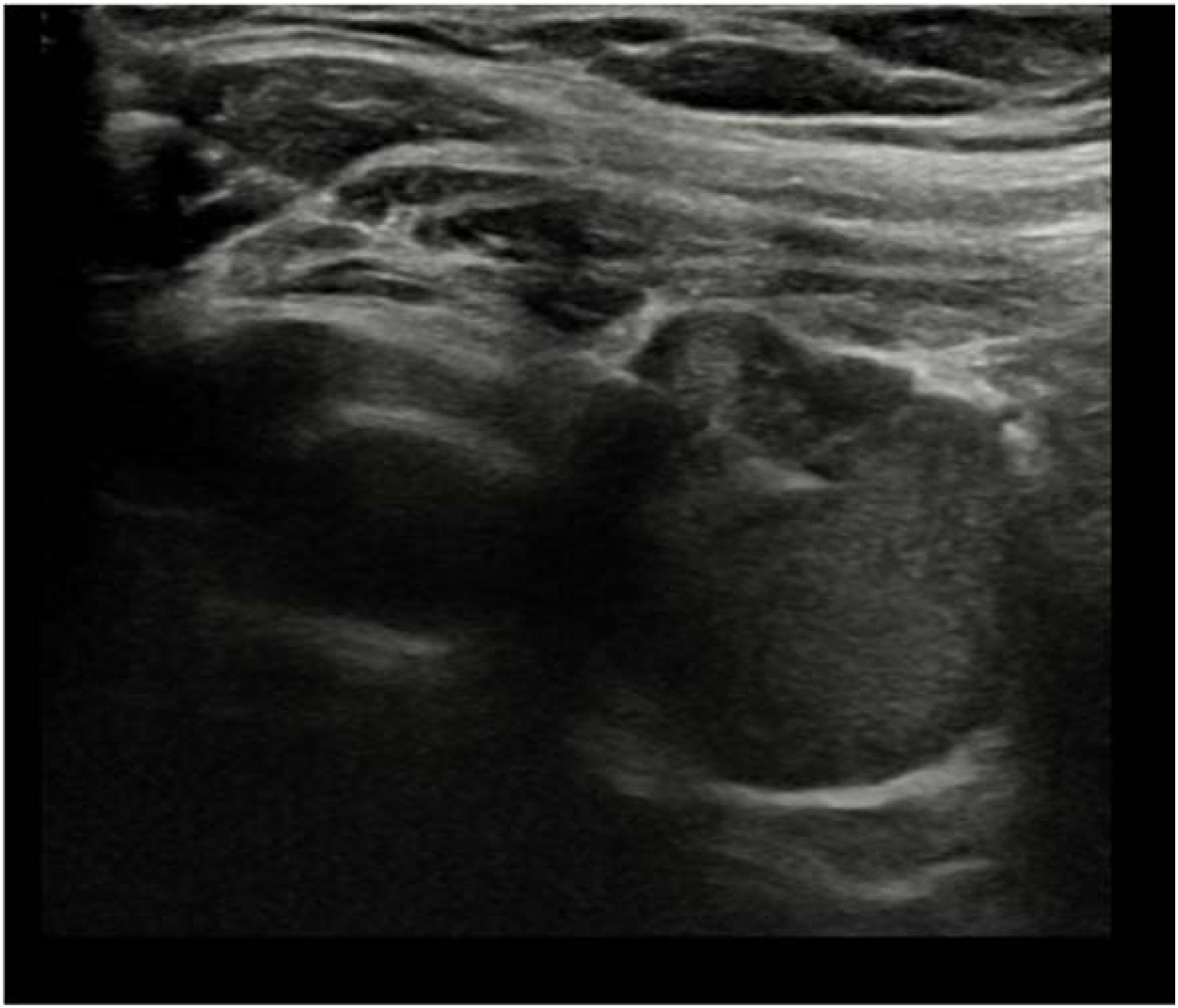
On cervical Ultrasound (US), a solid nodule measuring 1.1 × 0.5 × 0.5 cm was found in the left thyroid lobe, categorized as TI-RADS 3. The US was repeated, revealing an additional new nodule in the right thyroid lobe measuring 1.9 × 1.7 × 1.6 cm.
Investigation continued with a Computed Tomography (CT) scan of the neck, which showed a contrast-enhancing nodular image measuring 1.6 cm in the region of the right inferior parathyroid. A Fine-Needle Aspiration Biopsy (FNAB) of the left thyroid nodule was classified as benign (BETHESDA II). Serum ionic Calcium (iCa) and Parathyroid Hormone (PTH) levels were within normal limits: iCa 1.16 mmol/L (RR: 1.15–1.35 mmol/L) and PTH 35.0 pg/mL (RR: 16–65 pg/m).
After nine months, the patient returned for follow-up. A new ultrasound revealed growth of both nodules. Right lobe nodule: TI-RADS 5, measuring 1.9 × 1.4 × 1.7 cm. Left lobe nodule: TI-RADS 3, measuring 1.1 × 0.6 × 1.1 cm. FNAB of the right nodule resulted in atypia of undertemined significance (BETHESDA III) and was repeated six months later, resulting in follicular neoplasm ou suspicious for a follicular neoplasm (BETHESDA IV). In this latter evaluation, based solely on celular morphology, it was not possible to reliably determine the histiogenesis of the lesion (thyroid or parathyroid, as suggested by the ultrassound examination). Therefore, it was not possible to determine whether the lesion was of thyroid or parathyroid origin. At this time, PTH remained normal (33.5 pg/mL – RR: 1–65 pg/m), and ionic calcium was slightly below the reference range (1.13 mmol/L – RR: 1.15–1.35 mmol/L). At this time our suspicion was of a thyroid nodule so a right hemithyroidectomy with cervical lymph node dissection was proposed.
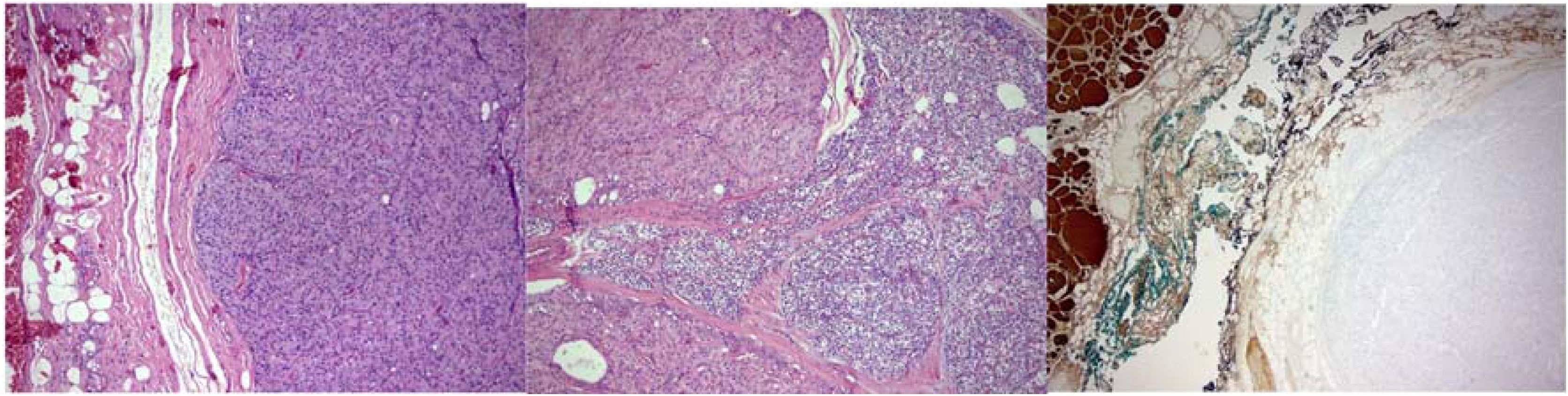
The specimen sent for histopathological examination consisted of a Bethesda IV right thyroid nodule and level VI lymph node dissection tissue. After a challenging pathological analysis and multiple slide revisions by three experienced pathologists, the final diagnosis was parathyroid carcinoma. Currently, the patient is under semiannual follow-up, with normal laboratory tests except for borderline low ionic calcium, and with no evidence of metastases (Figs. 1 and 2).


(A) Solid neoplastic proliferation composed of oncocytic cells with eosinophilic cytoplasm. (B) Area of capsular invasion and close contact with parathyroid tissue. (C) The lesion does not express thyroglobulin on immunohistochemistry (non-tumoral thyroid parenchyma positive as internal control on the left).
Scoping review.
Review questionsWhat is the clinical and laboratory profile of patients histopathologically diagnosed with intrathyroidal parathyroid carcinoma over the last 25-years? What are the diagnostic challenges?
Search strategyThe PubMed electronic database was used. Keywords included combinations of the following terms: (“Parathyroid carcinoma” OR “Carcinoma of parathyroid”) AND (“Intrathyroidal” OR “Intrathyroid” OR “Intrathyroidal parathyroid carcinoma” OR “Parathyroid carcinoma thyroid”).
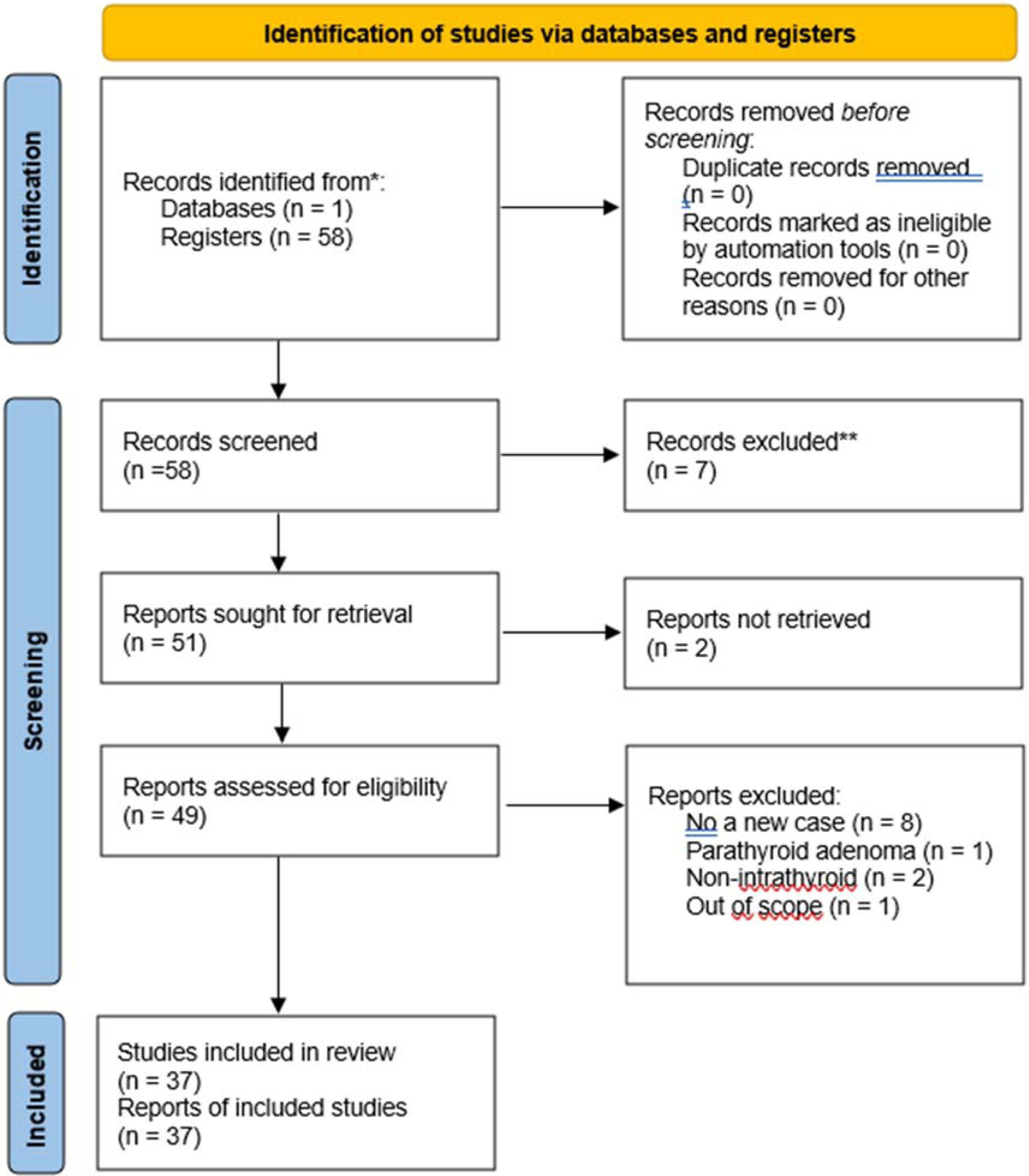
Selection processA total of 58 articles were identified, of which 7were excluded due to the time criteria and 2 were inaccessible. From the remaining 49 articles, 12 were excluded, leaving 37 articles to compose the scoping review. Including our case, the final number was 38 cases.
Inclusion criteriaStudies reporting a newly described, histologically confirmed case of intrathyroidal parathyroid carcinoma. Studies published within the last 25-years, from January 2000 to July 2025, that were accessible for full reading.
Exclusion criteriaStudies that did not report a new case of intrathyroidal parathyroid carcinoma. Studies with inappropriate methodology. Studies with insufficient information.
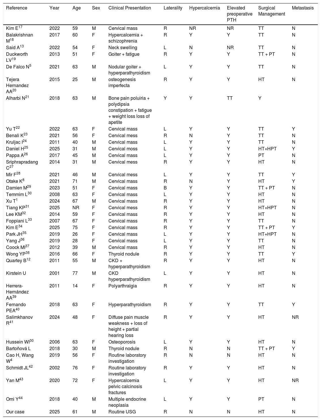
Data extractionData were extracted and tabulated to facilitate visualization of the following variables: author, year of publication, age, sex, lesion laterality, hypercalcemia, elevated PTH, surgical management, and presence of metastases (Fig. 3).
Discussion and conclusion
The results were discussed based on the existing evidence, considering clinical implications and recommendations for future practice.
The study limitations and areas for future research were also addressed.
ResultsA total of 51 articles were found in the PubMed database between 2000 and 2025 using the predefined descriptors. Of these, 2 were inaccessible and 12 were excluded according to the previously mentioned criteria, leaving 37 articles reporting previously unpublished cases of intrathyroidal parathyroid carcinoma.
DiscussionPrimary Hyperparathyroidism (PHPT) is an endocrine condition with a broad spectrum of clinical manifestations. Its main etiologies are adenoma, hyperplasia, or Parathyroid Carcinoma (PC). The latter is an especially rare endocrine neoplasm, accounting for only 0.005% of all malignancies.1
The parathyroid glands develop from the pharyngeal pouches, with the superior glands originating from the endoderm of the fourth pharyngeal pouch and the inferior glands from the endoderm of the third pharyngeal pouch, together with the thymus. They are composed of PTH-producing chief cells and oxyphil cells. Parathyroid glands are prone to ectopic locations due to aberrant migration during embryogenesis, occurring between the 5th and 6th weeks of gestation.2
The incidence of parathyroid ectopia ranges from 2% to 43% in the general population. When located within thyroid tissue, this percentage decreases to 1%–6%.1 In this context, a diseased parathyroid gland may be found inside the thyroid gland, such as Intrathyroidal Parathyroid Carcinoma (IPC), making this condition even rarer. Recent literature searches have identified approximately 37 cases reported over the last 25-years.
The etiology of parathyroid carcinoma remains largely unknown, although patients with familial hyperparathyroidism, Multiple Endocrine Neoplasia type 1 (MEN1), and those exposed to head and neck irradiation have an increased risk of developing the disease.3
Clinically, PC usually presents as a cervical mass and signs of severe hypercalcemia, with or without associated renal disease and metabolic bone disease. Diagnostic suspicion, already infrequent, becomes even more neglected since approximately 25% of patients present with few or only mild symptoms. Currently, around 2% of parathyroid cancers are detected through biochemical screening,3 and approximately 90% of these cases are hormonally active tumors, while the remaining are nonfunctioning.4,5
Nonfunctioning parathyroid carcinoma represents an exceptionally rare clinical entity, as most parathyroid carcinomas present with severe hyperparathyroidism and markedly elevated serum calcium and PTH levels. In contrast, nonfunctioning tumors are not associated with the characteristic biochemical abnormalities or functional imaging findings, such as sestamibi uptake, typically observed in primary hyperparathyroidism. Consequently, diagnosis may be particularly challenging and often relies primarily on histopathological and immunohistochemical evaluation. In addition, postoperative surveillance in these cases may be more complex, as biochemical monitoring has limited sensitivity for detecting recurrence. Therefore, follow-up should rely predominantly on imaging studies aimed at identifying local recurrence or distant metastases. Only a very limited number of comparable cases have been reported in the literature, some of which describe similar immunophenotypic findings, including negative PTH expression associated with positive galectin-3 staining.
The biochemical evaluation of suspected PHPT should include serum total calcium, PTH, creatinine, and 25-hydroxyvitamin D levels.6 In our case, the patient had no laboratory evidence of hyperparathyroidism, which did not prompt suspicion of parathyroid pathology. The first Fine-Needle Aspiration Biopsy (FNAB) of the right thyroid nodule showed atypia of undetermined significance result (BETHESDA III); in the second Fine-Needle Aspiration (FNA) repeated six months later of the same thyroid nodule, only minimal evidence of parathyroid cells was found, and therefore hemithyroidectomy was chosen. Clinically, attention should be paid to signs and symptoms of renal manifestations such as nephrolithiasis or renal failure, fragility fractures and osteoporosis, as well as neuropsychiatric, cognitive, musculoskeletal, and gastrointestinal complaints.6
The diagnosis of PC should be considered in the following situations: (a) Serum calcium >14 mg/dL; (b) Serum PTH levels >10 times the upper limit of normal; (c) Rapid onset of acute symptoms; (d) Detection of metastases on imaging studies.5
Imaging studies are useful, and cervical ultrasound remains the first-line imaging modality.1 Combining it with [99mTc]Tc-MIBI SPECT/CT is a widely available and accepted first-line strategy. PET/CT may be considered as an alternative method, and other radiopharmaceutical options may also be used. Four-Dimensional Computed Tomography (4D-CT) is useful in cases with negative or inconclusive imaging, in patients with distorted cervical anatomy, or after unsuccessful surgery. Magnetic resonance imaging can also be used when first-line imaging is negative or inconclusive, or in pregnant patients. Invasive diagnostic procedures should remain a last resort.7
Fine-Needle Aspiration (FNAB) is not recommended in suspected PC due to the risk of tumor seeding and metastasis along the needle track.5 It is noteworthy that in the present case, the patient underwent two thyroid FNABs, one of the right nodule and one of the left.
In the literature, the most common location of IPC is the right thyroid lobe (55.26%), which is consistent with our patient. However, in this case the lesion was located in the upper right thyroid lobe, whereas in the literature, it is most commonly found in the lower portions.8,9
The treatment of choice for primary hyperparathyroidism and parathyroid carcinoma is currently surgical.10,11 The optimal surgical technique remains under debate in order to improve survival outcomes. Schulte et al., in a cohort study, demonstrated that initial oncological en bloc resection ‒ including removal of the parathyroid lesion along with surrounding adipose tissue, the ipsilateral thyroid lobe, and level VI lymph nodes, achieving microscopically negative margins (R0) ‒ provides better local control and disease-free survival when compared to conservative surgery or local excisions. In contrast, local recurrence occurred in approximately 50% of cases with positive margins, highlighting the significantly increased risk when clear margins were not achieved.11 Jonathan B. also added that the aforementioned en bloc resection is associated with a local recurrence rate of only 8% and a long-term survival rate of approximately 90%.3
When no pathological gland is identified during initial surgery, routine bilateral neck exploration is mandatory. If the findings remain uncertain, thyroid lobectomy with isthmectomy may be considered if a thyroid nodule is found near the suspected site of the pathological gland.12
In our case, the patient underwent right hemithyroidectomy with level VI lymph node dissection (due to a palpable enlarged lymph node), with preservation of the four parathyroid glands, since intraoperative findings did not raise suspicion of parathyroid involvement.
Adjuvant therapeutic options for reducing hypercalcemia include calcimimetics and bisphosphonates, which are first-line treatments for PHPT. Immunobiological agents, such as denosumab, may stabilize calcium levels in cases of refractory hypercalcemia.1 In the context of IPC, these therapies may improve hypercalcemia-related symptoms; however, persistent symptoms after tumor resection should raise suspicion of surgical failure. Radiotherapy and chemotherapy should not be routinely used and are reserved for palliative cases or for reducing the risk of local recurrence. Therefore, surgery remains the only potentially curative treatment.3,13
Surgical success is defined by maintenance of eucalcemia during the arbitrarily accepted interval of 6-months after surgery.6,10 Surgical failure may be related to initial treatment with simple parathyroidectomy only, presence of nodal or distant metastatic disease at diagnosis, and nonfunctioning parathyroid carcinoma. Early reoperation should be considered in patients diagnosed with parathyroid carcinoma after simple parathyroidectomy.3
Regarding lymph node management in PC, there is controversy in the literature regarding the need for prophylactic central neck dissection. Hsu et al. reviewed 22-years of publications and concluded that only tumors ≥3 cm predicted lymph node metastasis, which were 7.5-times more frequent than in tumors <3 cm. Therefore, prophylactic central neck dissection may be indicated only in tumors larger than 3 cm.14
The definitive diagnosis and differentiation between thyroid and parathyroid neoplasms within the thyroid gland is only possible after histopathological analysis. In most cases, the diagnosis of IPC was not even considered prior to histopathological evaluation, as in our case. Oxyphilic cells in parathyroid lesions are indistinguishable from Hürthle/oncocytic cells in thyroid diseases. This distinction becomes even more difficult, if not impossible, when the parathyroid lesion is located inside the thyroid gland and there is no relevant clinical or radiological information, as in the present case. Cytology alone is insufficient to differentiate adenoma from PC.15 Immunohistochemistry plays an important role by identifying the expression and localization of specific proteins.
From an immunohistochemical perspective, Uljanovs et al.15 suggested that the histogenesis of parathyroid tumors involves positivity for markers such as PTH, chromogranin A, vimentin, cytokeratins, and GATA3, and negativity for TTF-1, thyroglobulin, CD56, and LCA. The pathogenesis includes alterations in cyclin D1 expression, calcium sensing, tumor microenvironment, and the local immune landscape. Loss of parafibromin favors the diagnosis of carcinoma over benign parathyroid lesions and is associated with poorer prognosis. Additionally, PC is associated with loss of parafibromin expression, increased Ki-67 activity (>5%), loss of APC, p27, Bcl-2, MDM2, RB or E-cadherin expression, and expression of PGP9.5, galectin-3, hTERT, or aberrant p53.
The histopathological specimen from right hemithyroidectomy with level VI lymph node dissection underwent multiple reviews. The intrathyroidal lesion showed close relationship with the parathyroid gland and focal capsular invasion. It measured 1.9 × 1.5 × 1.5 cm and was located in the upper third of the right lobe.
The cytoarchitectural pattern consisted of oncocytic-appearing cells arranged within fibrous trabeculae, with minimal cellular atypia, inconspicuous mitoses, absence of necrosis, and no evidence of vascular, lymphatic, or perineural invasion. The adjacent thyroid tissue showed no significant pathological changes, and the surgical margins were free of neoplasm. The level VI lymph nodes consisted of adipose tissue with vascular congestion and thymic remnants showing signs of regression.
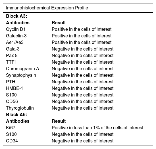
From a morphological standpoint, the lesion was more likely of parathyroid origin within the thyroid parenchyma. However, this could not be immunohistochemically confirmed due to lack of expression of parathyroid markers (PTH, GATA3, chromogranin, and synaptophysin). Considering the morphological features consistent with parathyroid cell histogenesis, the presence of focal capsular invasion raises the possibility of parathyroid carcinoma, possibly at an early stage, with preserved expression of galectin-3 and cyclin D1. However, in the absence of unequivocal invasive features such as vascular, lymphatic, or perineural invasion, the interpretation of capsular invasion should be approached with caution, particularly in intrathyroidal parathyroid lesions. The possibility of a thyroid-origin oncocytic lesion (adenoma or carcinoma) was initially excluded. Furthermore, exclusion of embryonic remnants should be performed by expanding the immunohistochemical panel to include CK5/6 and/or p63. In this context, the diagnostic criteria proposed by the 2022 WHO Classification of Parathyroid Tumors are particularly relevant.
According to the 2022 WHO Classification of Parathyroid Tumors,16 the diagnosis of parathyroid carcinoma requires unequivocal evidence of invasive growth, including vascular, lymphatic, or perineural invasion, direct invasion of adjacent structures, or the presence of metastatic disease. In contrast, lesions lacking these definitive criteria but presenting worrisome histopathological features are currently classified as atypical parathyroid tumors, representing neoplasms of uncertain malignant potential. In intrathyroidal parathyroid lesions, however, the interpretation of capsular or local invasion may be particularly challenging, as the gland may be partially embedded within the thyroid parenchyma or its pseudocapsule, potentially mimicking invasive growth.
Another noteworthy aspect of the present case is the unusual immunohistochemical profile. Parathyroid tumors typically retain immunoreactivity for Parathyroid Hormone (PTH) and GATA3, markers that help confirm parathyroid origin. Therefore, the absence of expression of these markers, although not entirely excluding parathyroid differentiation, represents an extremely uncommon immunophenotypic pattern. These findings highlight the diagnostic complexity of intrathyroidal parathyroid neoplasms and emphasize the importance of integrating histopathological, immunohistochemical, and clinical features when distinguishing parathyroid carcinoma from atypical parathyroid tumors.
Recent literature and the 2022 WHO Classification of Parathyroid Tumors16 recommend genetic evaluation of the CDC73 gene in patients diagnosed with parathyroid carcinoma, even in apparently sporadic cases, given the significant proportion of unrecognized germline mutations and its association with more aggressive disease behavior. In this context, assessment of parafibromin expression may also provide supportive diagnostic and prognostic information. However, in the present case, genetic testing was not performed due to lack of insurance coverage and the patient’s limited financial resources. Despite this limitation, the diagnosis was established based on the integration of histopathological findings, immunohistochemical profile, and clinical features.
The differential diagnosis between parathyroid and thyroid tumors, paragangliomas, hematological malignancies, or metastatic tumors can be clarified using PTH, chromogranin A, TTF-1, calcitonin, LCA, CD56, and vimentin, among other markers. Once again, immunohistochemistry proves to be an important tool in pathogenetic studies due to its ability to highlight both the presence and tissue localization of specific proteins.
The prognosis of parathyroid carcinoma remains poorly documented. However, it is evident that adequate surgical resection improves survival. IPC, due to its greater diagnostic difficulty, may delay definitive diagnosis and consequently decrease survival, although there is no concrete evidence that it is more lethal. The overall 10-year survival rate is estimated at 60–70%, due to its slow growth.1 Such patients should undergo regular surveillance with serum calcium and PTH measurements.3 Our patient remains under semiannual follow-up with recommended laboratory monitoring and has, so far, shown no evidence of metastases.
ConclusionParathyroid carcinoma accounts for approximately 1% of all cases of primary hyperparathyroidism and should be considered in the presence of markedly elevated PTH levels and severe hypercalcemia. Due to the frequent ectopic location of the parathyroid glands, malignant parathyroid disease may be found within the thyroid gland, making its incidence even lower and its diagnosis even more challenging, as in the case of Intrathyroidal Parathyroid Carcinoma (IPC).
After analyzing the IPC cases published over the last 25-years, summarized in Table 1, it was observed that the laboratory profile of patients involved hypercalcemia and elevated PTH levels in most cases (81.5%), compared to cases with no laboratory abnormalities (7.9%). Clinically, common presentations included cervical mass, goiter, bone pain, chronic kidney disease, and thyroid nodules detected during routine examination. The age ranged from 14- to 77-years, with a predominance of female patients, and the right side was the most commonly affected. The incidence of metastasis during follow-up was 23.6%.
Summary of reported intrathyroidal parathyroid carcinoma demographics, characteristics, and management.
| Reference | Year | Age | Sex | Clinical Presentation | Laterality | Hypercalcemia | Elevated preoperative PTH | Surgical Management | Metastasis |
|---|---|---|---|---|---|---|---|---|---|
| Kim E17 | 2022 | 59 | M | Cervical mass | R | NR | NR | TT | N |
| Balakrishnan M18 | 2017 | 60 | F | Hypercalcemia + schizophrenia | R | Y | Y | TT | N |
| Said A13 | 2022 | 54 | F | Neck swelling | L | N | NR | TT | N |
| Duckworth LV19 | 2013 | 51 | F | Goiter + fatigue | R | Y | Y | TT + PT | N |
| De Falco N5 | 2021 | 63 | M | Nodular goiter + hyperparathyroidism | L | Y | Y | TT | N |
| Tejera Hernandez AA20 | 2015 | 25 | M | osteogenesis imperfecta | R | Y | Y | HT | N |
| Alharbi N21 | 2018 | 63 | M | Bone pain poluiria + polydipsia constipation + fatigue + weight loss loss of apetite | Y | Y | TT | Y | |
| Yu T22 | 2022 | 63 | F | Cervical mass | L | Y | Y | TT | Y |
| Benali K23 | 2021 | 56 | F | Cervical mess | R | N | Y | TT | N |
| Kruljac I24 | 2011 | 40 | M | Cervical mass | L | Y | Y | TT | N |
| Daniel H25 | 2025 | 31 | M | Cervical moss | L | Y | Y | HT+HPT | Y |
| Pappa A26 | 2017 | 45 | M | Cervical mass | L | Y | Y | PT | N |
| Sriphrapradang C27 | 2014 | 31 | M | Cervical mess | R | Y | Y | HT | N |
| Mir F28 | 2021 | 46 | M | Cervical mess | L | Y | Y | TT | Y |
| Otake K8 | 2021 | 71 | M | Cervical mass | R | N | Y | HT | Y |
| Damien M29 | 2023 | 51 | F | Cervical mass | B | Y | Y | TT + PT | N |
| Temmim L30 | 2008 | 63 | F | Cervical mass | L | Y | Y | HT | N |
| Xu T1 | 2024 | 67 | M | Cervical mass | R | Y | Y | HT | N |
| Tiang KP31 | 2025 | NR | F | Cervical mess | R | Y | Y | HT+HPT | N |
| Lee KM32 | 2014 | 59 | F | Cervical mass | R | Y | Y | HT | N |
| Foppiani L33 | 2007 | 67 | F | Cervical mass | R | Y | Y | TT | N |
| Kim E34 | 2025 | 75 | F | Cervical mass | R | Y | Y | TT + PT | Y |
| Park JH35 | 2019 | 26 | F | Cervical mass | L | Y | Y | HT+HPT | N |
| Yang J36 | 2019 | 28 | F | Cervical mass | L | Y | Y | TT | N |
| Coock MI37 | 2012 | 39 | M | Cervical mass | R | Y | Y | HT | N |
| Wong YP38 | 2016 | 66 | F | Thyroid nodule | R | Y | Y | TT | Y |
| Quartey B12 | 2011 | 55 | M | CKD + hyperparathyroidism | R | Y | Y | HT | N |
| Kirstein U | 2001 | 77 | M | CKD hyperparathyroidism | L | Y | Y | HT | N |
| Herrera-Hernández AA39 | 2011 | 14 | F | Polyarthralgia | R | Y | Y | HT | N |
| Fernando PEA40 | 2018 | 63 | F | Hyperparathyroidism | R | Y | Y | TT | Y |
| Salimkhanov R41 | 2024 | 48 | F | Diffuse pain muscle weakness + loss of height + partial hearing loss | R | Y | Y | HT | NR |
| Hussein WI30 | 2006 | 63 | F | Osteoporosis | L | Y | Y | HT | N |
| Bartoňová L | 2018 | 30 | M | Thyroid nodule | R | N | N | TT + PT | Y |
| Cao H, Wang W4 | 2019 | 56 | F | Routine laboratory investigation | R | N | N | HT | N |
| Schmidt JL42 | 2002 | 76 | F | Routine laboratory investigation | R | Y | Y | HT | N |
| Yan M43 | 2020 | 72 | F | Hypercalcemia pelvic calcinosis fractures | L | Y | Y | HT | NR |
| Omi Y44 | 2018 | 40 | M | Multiple endocrine neoplasia | L | Y | Y | PT | N |
| Our case | 2025 | 61 | M | Routine USG | R | N | N | HT | N |
HT, Hemithyroidectomy; TT, Total Thyroidectomy; PT, Parathyroidectomy; NR, Not Reported.
Complete surgical resection is the gold standard treatment for parathyroid carcinoma and involves en bloc total tumor resection, avoiding capsular rupture, including surrounding adipose tissue, ipsilateral thyroidectomy, and level VI lymph node dissection. This approach provides better local disease control compared to conservative surgery or local excisions. When abnormal intraoperative appearance or suspected invasion of surrounding tissues is identified, the extent of resection should be expanded to include all involved tissue. It is likely that the diagnosis of IPC is only considered after histopathological analysis, making it most often a retrospective diagnosis.
Immunihistochemial expression profile in the specimen of interest.
| Immunohistochemical Expression Profile | |
|---|---|
| Block A3: | |
| Antibodies | Result |
| Cyclin D1 | Positive in the cells of interest |
| Galectin-3 | Positive in the cells of interest |
| Ae1/Ae3 | Positive in the cells of interest |
| Gata-3 | Negative in the cells of interest |
| Pax 8 | Negative in the cells of interest |
| TTF1 | Negative in the cells of interest |
| Chromogranin A | Negative in the cells of interest |
| Synaptophysin | Negative in the cells of interest |
| PTH | Negative in the cells of interest |
| HMBE-1 | Negative in the cells of interest |
| S100 | Negative in the cells of interest |
| CD56 | Negative in the cells of interest |
| Thyroglobulin | Negative in the cells of interest |
| Block A6: | |
| Antibodies | Result |
| Ki67 | Positive in less than 1% of the cells of interest |
| S100 | Negative in the cells of interest |
| CD34 | Negative in the cells of interest |
It is therefore concluded that improving survival in IPC depends on raising clinical suspicion. For this reason, and considering that many patients are asymptomatic, diagnosis remains a major challenge. Further investigation should always be pursued in the presence of any suspicion of malignancy, atypical cases of hyperparathyroidism, symptomatic thyroid nodules, or persistence of clinical hyperparathyroidism after surgical resection.
ORCID IDBeatriz Ferreira Maraccini: 0009-0005-0419-9111
Barbara Guidolin Froner: 0009-0003-6820-2493
Fernando Canola Alliegro: 0000-0002-1921-8558
Pablo Soares Gomes Pereira: 0009-0003-3327-1755
Leandro Luiz Lopes de Freitas: 0000-0002-5869-0791
Camila Matsunaga de Angellis: 0000-0001-9383-5506
Ana Maria da Cunha Mercante: 0000-0003-2340-3382
FundingThis research received no external funding and was self-financed by the authors.
Data availability statementThe authors declare that all data are available in repository.
The authors declare no conflicts of interest.
We thank all co-authors for their valuable contributions and the patient for allowing us to study and report this case.




