Journal Information
Vol. 90. Issue 4. (In progress)
(July - August 2024)
Share
Share
Download PDF
More article options
Visits
181
Vol. 90. Issue 4. (In progress)
(July - August 2024)
Letter to the editor
Full text access
Ultrasonography in Bell’s palsy: the elephant in the room
Visits
181
Ahmad J. Abdulsalama,b,
Corresponding author
dr.ahmad.j.abdulsalam@gmail.com

Corresponding author.
, Ameerah Alsaqobic, Murat Karaa
a Hacettepe University Medical School, Department of Physical and Rehabilitation Medicine, Ankara, Turkey
b Mubarak Alkabeer Hospital, Department of Physical Medicine and Rehabilitation, Jabriya, Kuwait
c Physical Medicine and Rehabilitation Hospital, Department of Physical Medicine and Rehabilitation, Andalous, Kuwait
This item has received
Article information
Full Text
Bibliography
Download PDF
Statistics
Figures (1)
Full Text
Dear Editor,

We read with interest the recently published article “Task force of the Brazilian Society of Otology — evaluation and management of peripheral facial palsy” by Pauna et al.1 We would like to congratulate the authors for their in-depth evidence-based review on the evaluation and management of peripheral facial palsy. However, we believe the utility of Ultrasound (US) in Bell’s palsy was overlooked.

The US imaging to assess peripheral nerve disorders is well established and has become one of the most useful modalities.2 Its diagnostic novelty and importance are sometimes being compared to electrodiagnostic studies.3 This is particularly true in peripheral nerve entrapment. Although electrodiagnostic tests are widely used, they are insufficient in detecting the exact location of nerve injury or the underlying cause. Moreover, morphological assessment of the nerve and surrounding structures is not possible.

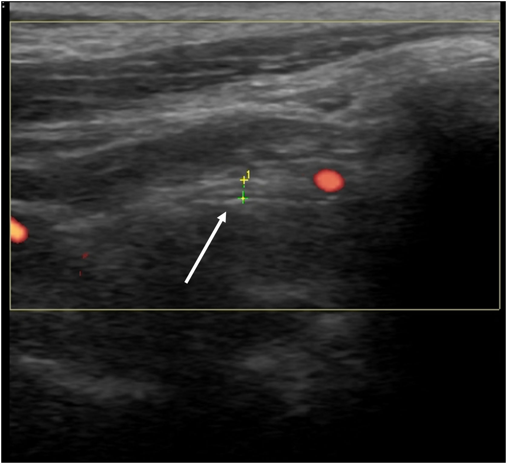
From a diagnostic perspective, US may provide evidence of nerve edema spreading distally from the site of facial nerve damage, making it a useful tool for evaluating the main trunk of the facial nerve in Bell’s palsy (Fig. 1).3 The prognostic value of US in this condition has also been highlighted in the literature.3 In one study, US was found superior to electrodiagnostic tests in predicting good versus poor recovery at three months post onset of weakness.4 The direct evaluation offered by US is less time consuming and efficient, in contrast to other modalities that are commonly used during evaluation and follow-up of Bell’s palsy.

Figure 1.

Ultrasound imaging/measurement (longitudinal view) of the facial nerve (white arrow) as they exit the stylomastoid foramen.

(0.13MB).

The utility of US in Bell’s palsy also extends to treatment. The validity as well as the reliability of US use in the management of facial nerve pathologies including Bell’s palsy has been documented in recent studies.4,5 To conclude, we underscore the potential role of US examination in Bell’s palsy in two ways. First, US can confirm the presence of an inflamed facial nerve and rule out any local pathology. Secondly, the exact site for injection can be localized. This in turn helps reduce the required blood drug concentration of corticosteroids and contributes to better recovery and less side effects.4

Conflicts of interest

The authors declare no conflicts of interest.

Ethical statement

Not applicable.

Disclosures/funding information/acknowledgments

None.

References
[1]
H.F. Pauna, V.A.R. Silva, J. Lavinsky, M.A. Hyppolito, M.F. Vianna, M.C.L. Gouveia, et al.
Task force of the Brazilian Society of Otology — evaluation and management of peripheral facial palsy.
Braz J Otorhinolaryngol, 90 (2023), pp. 101374
[2]
V. Ricci, C. Ricci, G. Cocco, F. Gervasoni, D. Donati, G. Farì, et al.
Histopathology and high-resolution ultrasound imaging for peripheral nerve (injuries).
J Neurol, 269 (2022), pp. 3663-3675
[3]
Y.L. Lo, S. Fook-Chong, T.H. Leoh, Y.F. Dan, M.P. Lee, H.Y. Gan, et al.
High-resolution ultrasound in the evaluation and prognosis of Bell’s palsy.
Eur J Neurol, 17 (2010), pp. 885-889
[4]
S.-H. Baek, Y.H. Kim, Y.-J. Kwon, J.H. Sung, M.H. Son, J.H. Lee, et al.
The utility of facial nerve ultrasonography in Bell’s palsy.
Otolaryngol Head Neck Surg, 162 (2020), pp. 186-192
[5]
S. Chhabda, D.S. Leger, R.K. Lingam.
Imaging the facial nerve: a contemporary review of anatomy and pathology.
Eur J Radiol, 126 (2020), pp. 108920
Copyright © 2024. Associação Brasileira de Otorrinolaringologia e Cirurgia Cérvico-Facial
Idiomas
Brazilian Journal of Otorhinolaryngology (English Edition)
Article options
Tools
en pt
Announcement Nota importante
Articles submitted as of May 1, 2022, which are accepted for publication will be subject to a fee (Article Publishing Charge, APC) payment by the author or research funder to cover the costs associated with publication. By submitting the manuscript to this journal, the authors agree to these terms. All manuscripts must be submitted in English.. Os artigos submetidos a partir de 1º de maio de 2022, que forem aceitos para publicação estarão sujeitos a uma taxa (Article Publishing Charge, APC) a ser paga pelo autor para cobrir os custos associados à publicação. Ao submeterem o manuscrito a esta revista, os autores concordam com esses termos. Todos os manuscritos devem ser submetidos em inglês.