
There appears to be no relationship between the size of tympanic perforations and hearing loss. Some studies in the literature have assessed this connection, with conflicting data and without proper methodology, especially concerning the measurement of the size of the perforation, which was performed in a subjective manner.
ObjectiveTo evaluate the size of tympanic perforations and to relate them to hearing loss in four different sound frequencies through the use of an objective method.
MethodsTransversal retrospective study. The present study evaluated 187 perforations through digital imaging, calculated the percentages of the tympanic membrane that was perforated using ImageScope software version 11.1.2.760 and correlated perforations’ size with hearing loss at four frequencies.
ResultsData were statistically analyzed using Pearson's correlation test.
ConclusionThere was no significant relationship between the size of tympanic perforations and hearing loss in the four analyzed frequencies.
Parece não haver relação entre o tamanho das perfurações timpânicas e a perda auditiva. Alguns trabalhos na literatura estudaram esta relação, com dados conflitantes e sem uso adequado da metodologia empregada, principalmente quanto à medição do tamanho da perfuração que se faz de modo subjetivo.
ObjetivoAnalisar através de um método objetivo o tamanho dessas perfurações e relacioná-las com perdas auditivas em quatro frequências sonoras.
MétodoEstudo retrospectivo de corte transversal. Foram avaliadas 187 perfurações timpânicas através de digitalização de imagem, medidas porcentualmente com o uso do software ImageScope Version 11.1.2.760 e correlacionadas com os limiares auditivos em quatro frequências.
ResultadosOs dados foram avaliados estatisticamente pelo teste de correlação de Pearson, que não demonstrou correlação entre o tamanho da perfuração timpânica e o grau de perda auditiva.
ConclusãoNão há relação significativa entre o tamanho das perfurações timpânicas e as quatro frequências estudadas.
This was a longitudinal retrospective cohort study. It is clear that there appears to be no direct relationship between the size of the tympanic membrane in simple chronic otitis media and hearing loss assessed by pure tone audiometry. This suspicion has been studied and evaluated, but by using subjective methods to measure the size of the perforations.1–5 With the advent of modern computer programs, the percentage of these perforations in relation to the total area of the membrane can be objectively evaluated. These more accurate data can be used to compare more reliably this finding with each audiometry frequency. Few similar studies were retrieved in the literature.6,7 This study aimed to analyze the correlation between the percentual size of the perforation and hearing loss in four frequencies.
MethodsThis was a retrospective cohort trial conducted at the Department of Otorhinolaryngology of a medical teaching institution, approved by the Research Ethics Committee under N° 9228. Images of the tympanic membrane were acquired using a 3mm diameter rigid fiber optic telescope coupled to a digital camera and with computer digital capture.
Only pictures of simple chronic otitis media (dry perforations as sequelae of necrotizing otitis) were selected, with more than six months without otorrhea reported by the patients. Hearing loss in four frequencies (500Hz, 1kHz, 2kHz, and 4kHz), with any degree of conductive hearing loss, was considered. The audiometries were performed by phonoaudiologists, using the Katz technique.8
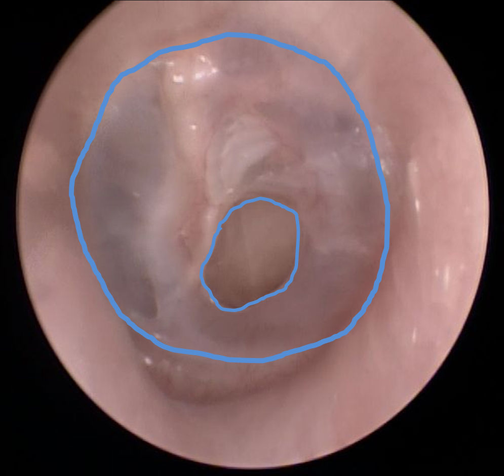
ImageScope, version 11.1.2.760 by Aperio Technologies®, was used. The selected images were evaluated by circumscribing (by tracking with a mouse) the total area of the tympanic membrane, which was then measured by pixel counting (Fig. 1). The same procedure was applied to the area of the perforation. Both measures were transported to an Excel® (Microsoft) spreadsheet. Since the determination of the area of perforation was then calculated as a percentage of the area of the tympanic membrane, there was no distortion because of the angle of view or the proximity of the image capture. These measurements were performed by two examiners, at different times, and only those that coincided with an error factor of <5% were considered.

The inclusion and exclusion criteria of the study were as follows:
- -
Inclusion – images of tympanic membrane with perforation without evidence of inflammation or otorrhea for more than six months duration, simple chronic otitis media.
- -
Exclusion – evaluation of the perforation size by two examiners presenting a difference >5%.
The audiograms were evaluated only with respect to their conductive hearing loss, i.e. the air-bone gap that characterized the tympanic involvement (membrane or ossicular chain). The following frequencies were used: 500Hz, 1000Hz, 2000Hz and 4000Hz.
Data obtained from the perforations of the tympanic membranes were correlated with the air-bone gap in each of the frequencies analyzed by Pearson's correlation test.
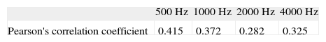
ResultsThe study included 187 ears that presented simple chronic otitis media. The age of patients analyzed ranged from 4 to 75 years. The right ear was involved in 79 patients, while 108 exhibited the problem in left ear. The correlation between the size of the perforation and the frequencies is listed in Table 1.
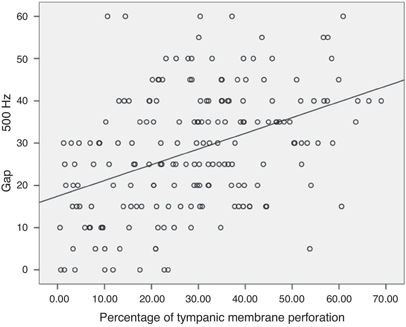
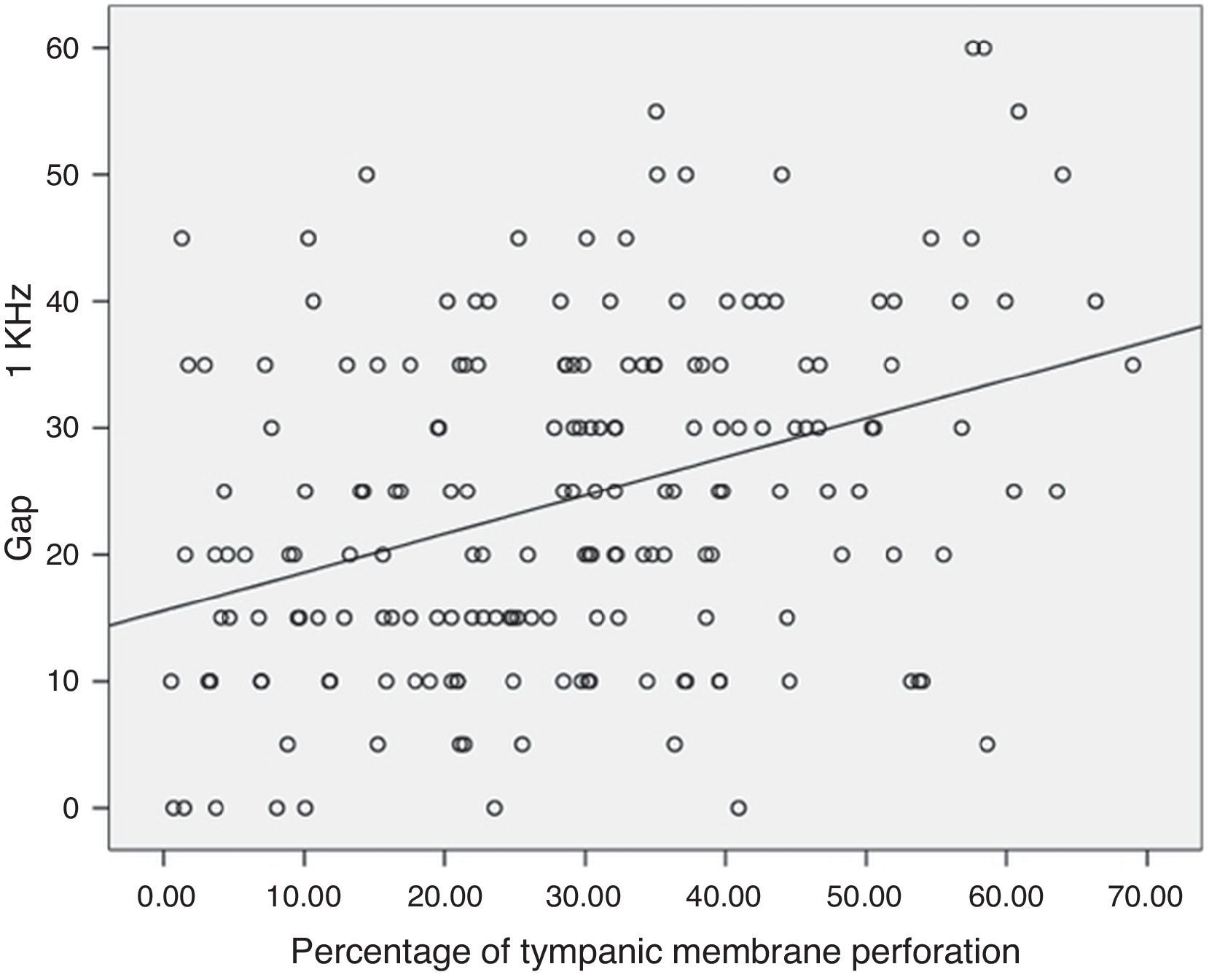
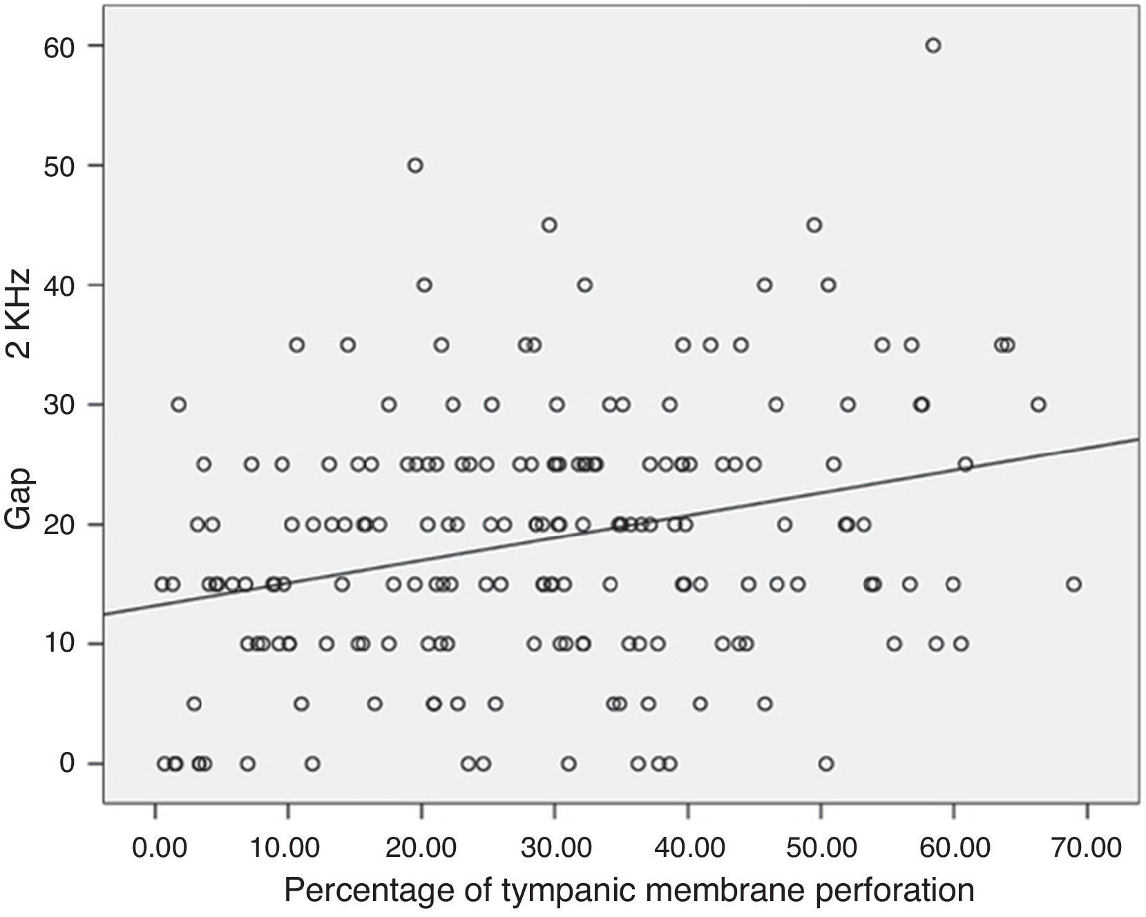
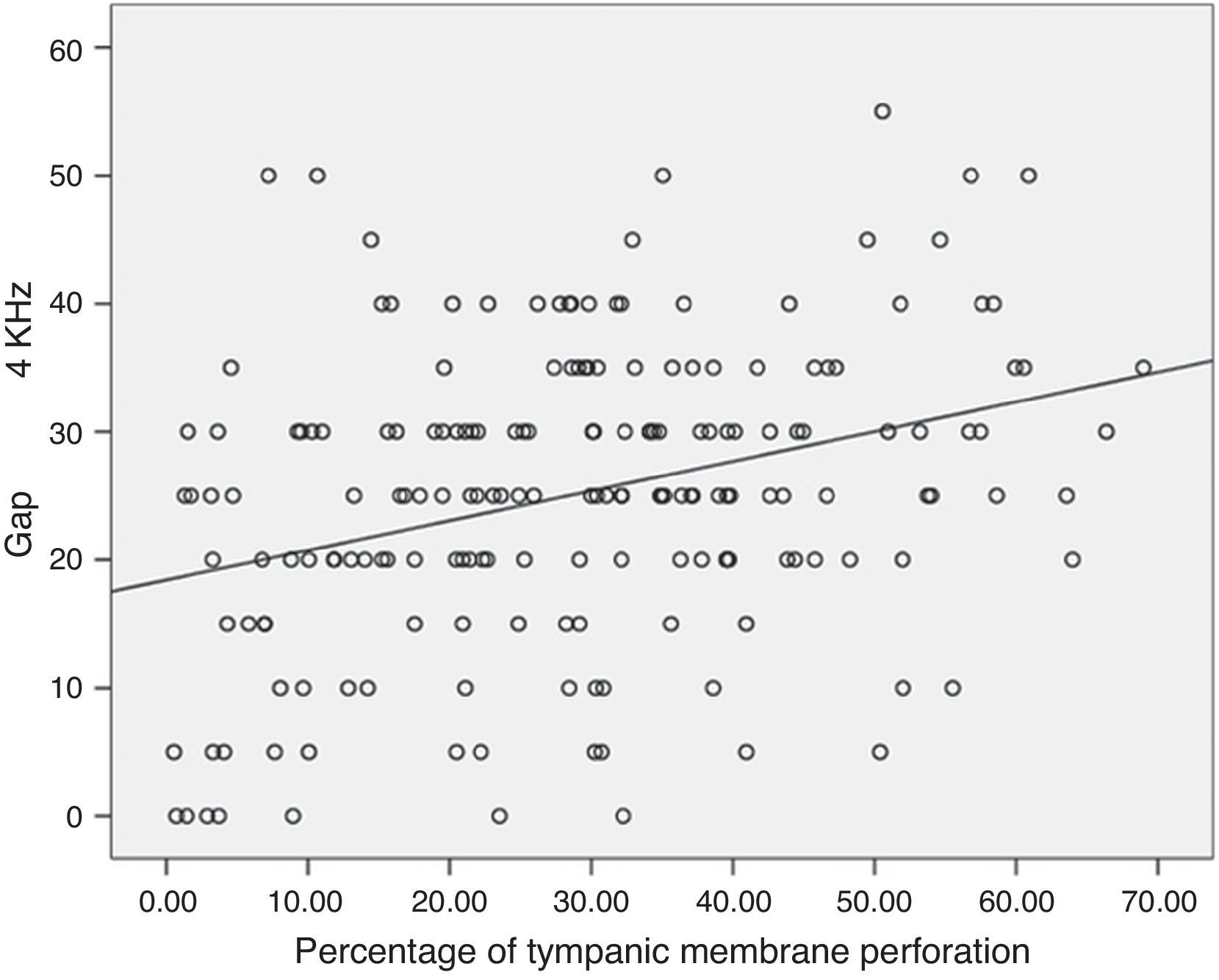
The correlation between the percentage of perforation of the tympanic membrane and the air-bone gap that exists in each of the evaluated frequencies is shown in Figs. 2–5.


Fig. 2 illustrates the correlation between the percentage of perforation of the tympanic membrane and the air-bone gap observed at a frequency of 500Hz.
Fig. 3 shows the correlation between the percentage of perforation of the tympanic membrane and the air-bone gap observed at a frequency of 1000Hz.
In Fig. 4, a correlation between the percentage of perforation of the tympanic membrane and the air-bone gap observed in the frequency of 2000Hz is shown.
Finally, in Fig. 5, the correlation between the percentage of perforation of the tympanic membrane and the air-bone gap observed at a frequency of 4000Hz is shown.
DiscussionThe linear correlation between the size of the tympanic perforation in patients with simple chronic otitis media and hearing loss in four different frequencies was investigated. Pearson's correlation coefficient for the frequencies of 500Hz, 1000Hz, 2000Hz and 4000Hz was, respectively, 0.415, 0.372, 0.282 and 0.325, demonstrating that there is a strong linear correlation between the variables studied.
The correlation for the frequency of 500Hz was found to be moderately significant for the issue examined, while the correlations observed for the other frequencies proved to be of little significance.
In the literature, Pannu et al.9 reported different results, demonstrating an increase in hearing loss with increasing sizes of the tympanic perforation in 100 patients who also had perforations without signs of active inflammation or secretion. Importantly, in that study, the perforation size was estimated by measuring their greater vertical (R1) and greater horizontal (R2) diameters with a 1-mm wire, inserting the values into the formula: perforation area=π×R1×R2.
Ibekwe et al.,10 analyzed 67 patients with a total of 77 perforations. Using the Pearson correlation: p=0.01, r=0.05, they concluded that the larger the tympanic membrane perforation, the greater the loss in sound perception.
The article by Ahmad and Ramani1 is in agreement with the studies previously mentioned. In that study, 70 patients with dry central perforation were analyzed. The patients were divided into four groups according to the size, expressed as a percentage, of the observed perforation. These authors analyzed the hearing loss in each frequency in each of their groups, and concluded that the hearing loss increased with the size of perforation.
Therefore, the present study, with a larger number of patients and using a more modern methodology, contradicts the literature and leads to the conclusion that factors, other than the size of perforation (e.g., disjunctions or fixations of the ossicular chain), compromise the auditory acuity in patients with simple chronic otitis media.
ConclusionThere was no correlation between the size of tympanic membrane perforations in simple chronic otitis media and hearing loss at 500Hz; 1000Hz; 2000Hz and 4000Hz.
Conflicts of interestThe authors declare no conflicts of interest.
Please cite this article as: Ribeiro FA, Gaudino VR, Pinheiro CD, Marçal GJ, Mitre EI. Objective comparison between perforation and hearing loss. Braz J Otorhinolaryngol. 2014;80:386–9.
Institution: Faculty of Medical Sciences, Santa Casa de São Paulo, São Paulo, SP, Brazil.
gology is pleased to honor the reviewers


